 Movie
Movie Controller

[English] 日本語
 Yorodumi
- PDB-3db1: Crystal structure of the 2H-phosphatase domain of Sts-2 in comple... -
+ Open data

- Basic information
| Entry | Database: PDB / ID: 3db1 | ||||||
|---|---|---|---|---|---|---|---|
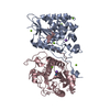
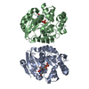
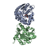
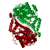
| Title | Crystal structure of the 2H-phosphatase domain of Sts-2 in complex with phosphate | ||||||
 Components Components | STS-2 protein | ||||||
 Keywords Keywords | HYDROLASE / Sts-2 / 2H-phosphatase / PGM domain / phosphate / SH3 domain | ||||||
| Function / homology |  Function and homology information Function and homology informationnegative regulation of immune system process / negative regulation of T cell receptor signaling pathway / regulation of cytokine production / nuclear speck / nucleoplasm / nucleus / cytoplasm / cytosol Similarity search - Function | ||||||
| Biological species |  | ||||||
| Method |  X-RAY DIFFRACTION / SYNCHROTRON / FOURIER SYNTHESIS / Resolution: 2.77 Å X-RAY DIFFRACTION / SYNCHROTRON / FOURIER SYNTHESIS / Resolution: 2.77 Å | ||||||
 Authors Authors | Nassar, N. / Chen, Y. / Carpino, N. | ||||||
 Citation Citation | Journal: Biochemistry / Year: 2009 Title: Structural and functional characterization of the 2H-phosphatase domain of Sts-2 reveals an acid-dependent phosphatase activity. Authors: Chen, Y. / Jakoncic, J. / Carpino, N. / Nassar, N. | ||||||
| History |
|
- Structure visualization
| Structure viewer | Molecule: MolmilJmol/JSmol |
|---|
- Downloads & links
-Download
| PDBx/mmCIF format | 3db1.cif.gz | 215 KB | Display | PDBx/mmCIF format |
|---|---|---|---|---|
| PDB format | pdb3db1.ent.gz | 175.2 KB | Display | PDB format |
| PDBx/mmJSON format | 3db1.json.gz | Tree view | PDBx/mmJSON format | |
| Others |  Other downloads Other downloads |
-Validation report
| Arichive directory | https://data.pdbj.org/pub/pdb/validation_reports/db/3db1ftp://data.pdbj.org/pub/pdb/validation_reports/db/3db1 | HTTPS FTP |
|---|
-Related structure data
| Related structure data | 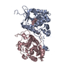 3d4iSC  3d6aC S: Starting model for refinement C: citing same article ( |
|---|---|
| Similar structure data |


-Links
 PDBj
PDBj


- Assembly
| Deposited unit | 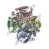
| |||||||||||||||||||||||||||||||||||||||||||||||||||||||||||||||||||||||||||||||||||||||||||||||||||||||||||||||||||||||||||||||||||||||||||||||||||||||||
|---|---|---|---|---|---|---|---|---|---|---|---|---|---|---|---|---|---|---|---|---|---|---|---|---|---|---|---|---|---|---|---|---|---|---|---|---|---|---|---|---|---|---|---|---|---|---|---|---|---|---|---|---|---|---|---|---|---|---|---|---|---|---|---|---|---|---|---|---|---|---|---|---|---|---|---|---|---|---|---|---|---|---|---|---|---|---|---|---|---|---|---|---|---|---|---|---|---|---|---|---|---|---|---|---|---|---|---|---|---|---|---|---|---|---|---|---|---|---|---|---|---|---|---|---|---|---|---|---|---|---|---|---|---|---|---|---|---|---|---|---|---|---|---|---|---|---|---|---|---|---|---|---|---|---|
| 1 | 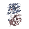
| |||||||||||||||||||||||||||||||||||||||||||||||||||||||||||||||||||||||||||||||||||||||||||||||||||||||||||||||||||||||||||||||||||||||||||||||||||||||||
| 2 | 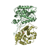
| |||||||||||||||||||||||||||||||||||||||||||||||||||||||||||||||||||||||||||||||||||||||||||||||||||||||||||||||||||||||||||||||||||||||||||||||||||||||||
| Unit cell |
| |||||||||||||||||||||||||||||||||||||||||||||||||||||||||||||||||||||||||||||||||||||||||||||||||||||||||||||||||||||||||||||||||||||||||||||||||||||||||
| Noncrystallographic symmetry (NCS) | NCS domain:
NCS domain segments: Ens-ID: 1 / Refine code: 1
|