 Movie
Movie Controller

[English] 日本語
 Yorodumi
- PDB-1m8k: Crystal Structure Of Methanobacterium Thermoautotrophicum Nicotin... -
+ Open data

- Basic information
| Entry | Database: PDB / ID: 1m8k | ||||||
|---|---|---|---|---|---|---|---|
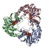
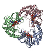
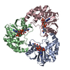
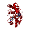
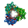
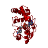
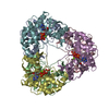
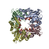
| Title | Crystal Structure Of Methanobacterium Thermoautotrophicum Nicotinamide Mononucleotide Adenylyltransferase Mutant H19A complexed with NAD | ||||||
 Components Components | Nicotinamide-nucleotide Adenylyltransferase | ||||||
 Keywords Keywords | TRANSFERASE / nucleotidyltransferase HXGH active site motif | ||||||
| Function / homology |  Function and homology information Function and homology informationnicotinamide-nucleotide adenylyltransferase / nicotinamide-nucleotide adenylyltransferase activity / NAD+ biosynthetic process / ATP binding / cytoplasm Similarity search - Function | ||||||
| Biological species |   Methanothermobacter thermautotrophicus (archaea) Methanothermobacter thermautotrophicus (archaea) | ||||||
| Method |  X-RAY DIFFRACTION / MOLECULAR REPLACEMENT / Resolution: 3 Å X-RAY DIFFRACTION / MOLECULAR REPLACEMENT / Resolution: 3 Å | ||||||
 Authors Authors | Saridakis, V. / Pai, E.F. | ||||||
 Citation Citation | Journal: J.Biol.Chem. / Year: 2003 Title: Mutational, Structural, and Kinetic Studies of the ATP-binding Site of Methanobacterium thermoautotrophicum Nicotinamide Mononucleotide Adenylyltransferase Authors: Saridakis, V. / Pai, E.F. | ||||||
| History |
|
- Structure visualization
| Structure viewer | Molecule: MolmilJmol/JSmol |
|---|
- Downloads & links
-Download
| PDBx/mmCIF format | 1m8k.cif.gz | 113.8 KB | Display | PDBx/mmCIF format |
|---|---|---|---|---|
| PDB format | pdb1m8k.ent.gz | 90.8 KB | Display | PDB format |
| PDBx/mmJSON format | 1m8k.json.gz | Tree view | PDBx/mmJSON format | |
| Others |  Other downloads Other downloads |
-Validation report
| Arichive directory | https://data.pdbj.org/pub/pdb/validation_reports/m8/1m8kftp://data.pdbj.org/pub/pdb/validation_reports/m8/1m8k | HTTPS FTP |
|---|
-Related structure data
| Related structure data |  1m8fC  1m8gC  1m8jC  1ej2S C: citing same article ( S: Starting model for refinement |
|---|---|
| Similar structure data |
-Links
 PDBj
PDBj- Assembly
| Deposited unit | 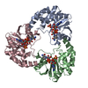
| ||||||||
|---|---|---|---|---|---|---|---|---|---|
| 1 | 
| ||||||||
| 2 |
| ||||||||
| Unit cell |
| ||||||||
| Details | The protein is made up of a dimer of trimers. |
-Components
| #1: Protein | Mass: 20497.660 Da / Num. of mol.: 3 / Mutation: H19A Source method: isolated from a genetically manipulated source Source: (gene. exp.) Methanothermobacter thermautotrophicus (archaea)Gene: mth150 / Plasmid: pET15B / Species (production host): Escherichia coli / Production host:  References: UniProt: O26253, nicotinamide-nucleotide adenylyltransferase #2: Chemical |   Mass: 96.063 Da / Num. of mol.: 3 / Source method: obtained synthetically / Formula: SO4 Mass: 96.063 Da / Num. of mol.: 3 / Source method: obtained synthetically / Formula: SO4#3: Chemical |   Mass: 663.425 Da / Num. of mol.: 3 / Source method: obtained synthetically / Formula: C21H27N7O14P2 / Comment: NAD*YM Mass: 663.425 Da / Num. of mol.: 3 / Source method: obtained synthetically / Formula: C21H27N7O14P2 / Comment: NAD*YM |
|---|
-Experimental details
-Experiment
| Experiment | Method: X-RAY DIFFRACTION / Number of used crystals: 1 |
|---|
- Sample preparation
| Crystal | Density Matthews: 4.07 Å3/Da / Density % sol: 69.74 % | ||||||||||||||||||||||||||||||||||||||||||||||||
|---|---|---|---|---|---|---|---|---|---|---|---|---|---|---|---|---|---|---|---|---|---|---|---|---|---|---|---|---|---|---|---|---|---|---|---|---|---|---|---|---|---|---|---|---|---|---|---|---|---|
| Crystal grow | Temperature: 298 K / Method: vapor diffusion, hanging drop / pH: 8 Details: 1.5 M Ammonium Sulfate, 100 mM Tris, pH 8, VAPOR DIFFUSION, HANGING DROP, temperature 298K | ||||||||||||||||||||||||||||||||||||||||||||||||
| Crystal grow | *PLUS Temperature: 20 ℃ / pH: 8 / Method: vapor diffusion, hanging drop | ||||||||||||||||||||||||||||||||||||||||||||||||
| Components of the solutions | *PLUS
|
-Data collection
| Diffraction | Mean temperature: 100 K |
|---|---|
| Diffraction source | Source: ROTATING ANODE / Type: RIGAKU RU300 / Wavelength: 1.54 Å |
| Detector | Type: MARRESEARCH / Detector: IMAGE PLATE / Date: Sep 18, 2001 |
| Radiation | Protocol: SINGLE WAVELENGTH / Monochromatic (M) / Laue (L): M / Scattering type: x-ray |
| Radiation wavelength | Wavelength: 1.54 Å / Relative weight: 1 |
| Reflection | Resolution: 3→15 Å / Num. obs: 20207 / % possible obs: 100 % / Observed criterion σ(I): -3 / Redundancy: 11 % / Rmerge(I) obs: 0.14 / Rsym value: 0.192 / Net I/σ(I): 20.6 |
| Reflection shell | Resolution: 3→3.11 Å / Rmerge(I) obs: 0.393 / Mean I/σ(I) obs: 6.5 / Num. unique all: 2012 / Rsym value: 0.418 |
| Reflection | *PLUS Highest resolution: 3 Å / Num. measured all: 223204 / Rmerge(I) obs: 0.14 |
| Reflection shell | *PLUS % possible obs: 100 % |
- Processing
| Software |
| ||||||||||||||||||||||||||||||||||||||||||||||||||||||||||||||||||||||||||||||||
|---|---|---|---|---|---|---|---|---|---|---|---|---|---|---|---|---|---|---|---|---|---|---|---|---|---|---|---|---|---|---|---|---|---|---|---|---|---|---|---|---|---|---|---|---|---|---|---|---|---|---|---|---|---|---|---|---|---|---|---|---|---|---|---|---|---|---|---|---|---|---|---|---|---|---|---|---|---|---|---|---|---|
| Refinement | Method to determine structure: MOLECULAR REPLACEMENT Starting model: pdb entry 1ej2 Resolution: 3→14.99 Å / Rfactor Rfree error: 0.005 / Data cutoff high absF: 317851.62 / Data cutoff high rms absF: 317851.62 / Data cutoff low absF: 0 / Isotropic thermal model: RESTRAINED / Cross valid method: THROUGHOUT / σ(F): 2
| ||||||||||||||||||||||||||||||||||||||||||||||||||||||||||||||||||||||||||||||||
| Solvent computation | Solvent model: FLAT MODEL / Bsol: 14.3492 Å2 / ksol: 0.342116 e/Å3 | ||||||||||||||||||||||||||||||||||||||||||||||||||||||||||||||||||||||||||||||||
| Displacement parameters | Biso mean: 28.4 Å2
| ||||||||||||||||||||||||||||||||||||||||||||||||||||||||||||||||||||||||||||||||
| Refine analyze |
| ||||||||||||||||||||||||||||||||||||||||||||||||||||||||||||||||||||||||||||||||
| Refinement step | Cycle: LAST / Resolution: 3→14.99 Å
| ||||||||||||||||||||||||||||||||||||||||||||||||||||||||||||||||||||||||||||||||
| Refine LS restraints |
| ||||||||||||||||||||||||||||||||||||||||||||||||||||||||||||||||||||||||||||||||
| LS refinement shell | Resolution: 3→3.19 Å / Rfactor Rfree error: 0.017 / Total num. of bins used: 6
| ||||||||||||||||||||||||||||||||||||||||||||||||||||||||||||||||||||||||||||||||
| Xplor file |
| ||||||||||||||||||||||||||||||||||||||||||||||||||||||||||||||||||||||||||||||||
| Refinement | *PLUS Lowest resolution: 15 Å | ||||||||||||||||||||||||||||||||||||||||||||||||||||||||||||||||||||||||||||||||
| Solvent computation | *PLUS | ||||||||||||||||||||||||||||||||||||||||||||||||||||||||||||||||||||||||||||||||
| Displacement parameters | *PLUS | ||||||||||||||||||||||||||||||||||||||||||||||||||||||||||||||||||||||||||||||||
| Refine LS restraints | *PLUS
|
