 Movie
Movie Controller

[English] 日本語
 Yorodumi
- PDB-1f9a: CRYSTAL STRUCTURE ANALYSIS OF NMN ADENYLYLTRANSFERASE FROM METHAN... -
+ Open data

- Basic information
| Entry | Database: PDB / ID: 1f9a | ||||||
|---|---|---|---|---|---|---|---|
| Title | CRYSTAL STRUCTURE ANALYSIS OF NMN ADENYLYLTRANSFERASE FROM METHANOCOCCUS JANNASCHII | ||||||
 Components Components | HYPOTHETICAL PROTEIN MJ0541 | ||||||
 Keywords Keywords | TRANSFERASE / alpha/beta / HYPOTHETICAL PROTEIN / STRUCTURAL GENOMICS | ||||||
| Function / homology |  Function and homology information Function and homology informationnicotinamide-nucleotide adenylyltransferase / nicotinamide-nucleotide adenylyltransferase activity / NAD+ biosynthetic process / ATP binding / cytoplasm Similarity search - Function | ||||||
| Biological species |   Methanocaldococcus jannaschii (archaea) Methanocaldococcus jannaschii (archaea) | ||||||
| Method |  X-RAY DIFFRACTION / SYNCHROTRON / SIR / Resolution: 2 Å X-RAY DIFFRACTION / SYNCHROTRON / SIR / Resolution: 2 Å | ||||||
 Authors Authors | D'Angelo, I. / Raffaelli, N. / Dabusti, V. / Lorenzi, T. / Magni, G. / Rizzi, M. | ||||||
 Citation Citation | Journal: Structure Fold.Des. / Year: 2000 Title: Structure of nicotinamide mononucleotide adenylyltransferase: a key enzyme in NAD(+) biosynthesis. Authors: D'Angelo, I. / Raffaelli, N. / Dabusti, V. / Lorenzi, T. / Magni, G. / Rizzi, M. #1: Journal: J.Bacteriol. / Year: 1997Title: Characterization of nicotinamide mononucleotide adenylyltransferase from Thermophilic archaea Authors: Raffaelli, N. / Pisani, F.M. / Lorenzi, T. / Emanuelli, M. / Amici, A. / Ruggieri, S. / Magni, G. | ||||||
| History |
|
- Structure visualization
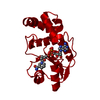
| Structure viewer | Molecule: MolmilJmol/JSmol |
|---|
- Downloads & links
-Download
| PDBx/mmCIF format | 1f9a.cif.gz | 230.3 KB | Display | PDBx/mmCIF format |
|---|---|---|---|---|
| PDB format | pdb1f9a.ent.gz | 184.7 KB | Display | PDB format |
| PDBx/mmJSON format | 1f9a.json.gz | Tree view | PDBx/mmJSON format | |
| Others |  Other downloads Other downloads |
-Validation report
| Arichive directory | https://data.pdbj.org/pub/pdb/validation_reports/f9/1f9aftp://data.pdbj.org/pub/pdb/validation_reports/f9/1f9a | HTTPS FTP |
|---|
-Related structure data
| Similar structure data |
|---|

-Links
 PDBj
PDBj- Assembly
| Deposited unit | 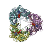
| ||||||||
|---|---|---|---|---|---|---|---|---|---|
| 1 |
| ||||||||
| Unit cell |
| ||||||||
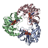
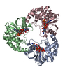
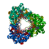
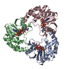
| Details | The asymmetric unit contains and homohexamer having 32 point group symmetry |
-Components
| #1: Protein | Mass: 19621.697 Da / Num. of mol.: 6 / Fragment: NMN ADENYLYLTRANSFERASE Source method: isolated from a genetically manipulated source Source: (gene. exp.) Methanocaldococcus jannaschii (archaea)Plasmid: PT7-7 / Production host:  #2: Chemical | ChemComp-MG /   Mass: 24.305 Da / Num. of mol.: 6 / Source method: obtained synthetically / Formula: Mg Mass: 24.305 Da / Num. of mol.: 6 / Source method: obtained synthetically / Formula: Mg#3: Chemical | ChemComp-ATP /   Mass: 507.181 Da / Num. of mol.: 6 / Source method: obtained synthetically / Formula: C10H16N5O13P3 / Comment: ATP, energy-carrying molecule*YM Mass: 507.181 Da / Num. of mol.: 6 / Source method: obtained synthetically / Formula: C10H16N5O13P3 / Comment: ATP, energy-carrying molecule*YM#4: Water | ChemComp-HOH / |  Mass: 18.015 Da / Num. of mol.: 664 / Source method: isolated from a natural source / Formula: H2O Mass: 18.015 Da / Num. of mol.: 664 / Source method: isolated from a natural source / Formula: H2O |
|---|
-Experimental details
-Experiment
| Experiment | Method: X-RAY DIFFRACTION / Number of used crystals: 1 |
|---|
- Sample preparation
| Crystal | Density Matthews: 2.54 Å3/Da / Density % sol: 53 % | |||||||||||||||||||||||||||||||||||
|---|---|---|---|---|---|---|---|---|---|---|---|---|---|---|---|---|---|---|---|---|---|---|---|---|---|---|---|---|---|---|---|---|---|---|---|---|
| Crystal grow | Temperature: 277 K / Method: vapor diffusion, hanging drop / pH: 7.5 Details: Jeffamine M600, Hepes, cesium chloride, pH 7.5, VAPOR DIFFUSION, HANGING DROP, temperature 277K | |||||||||||||||||||||||||||||||||||
| Crystal grow | *PLUS Temperature: 4 ℃ | |||||||||||||||||||||||||||||||||||
| Components of the solutions | *PLUS
|
-Data collection
| Diffraction | Mean temperature: 100 K |
|---|---|
| Diffraction source | Source: SYNCHROTRON / Site: EMBL/DESY, HAMBURG  / Beamline: BW7B / Wavelength: 0.8139 / Beamline: BW7B / Wavelength: 0.8139 |
| Detector | Type: MARRESEARCH / Detector: IMAGE PLATE / Date: Mar 20, 1999 |
| Radiation | Protocol: SINGLE WAVELENGTH / Monochromatic (M) / Laue (L): M / Scattering type: x-ray |
| Radiation wavelength | Wavelength: 0.8139 Å / Relative weight: 1 |
| Reflection | Resolution: 2→20 Å / Num. all: 200580 / Num. obs: 80232 / % possible obs: 96 % / Observed criterion σ(F): 0 / Observed criterion σ(I): 2 / Redundancy: 2.5 % / Biso Wilson estimate: 25.5 Å2 / Rmerge(I) obs: 0.054 / Net I/σ(I): 8 |
| Reflection shell | Resolution: 2→20 Å / Redundancy: 2.2 % / Rmerge(I) obs: 0.178 / Num. unique all: 11668 / % possible all: 95.6 |
| Reflection | *PLUS % possible obs: 96 % / Num. measured all: 200580 |
- Processing
| Software |
| |||||||||||||||||||||||||
|---|---|---|---|---|---|---|---|---|---|---|---|---|---|---|---|---|---|---|---|---|---|---|---|---|---|---|
| Refinement | Method to determine structure: SIR / Resolution: 2→20 Å / σ(F): 0 / σ(I): 0 / Stereochemistry target values: Engh and Huber / Details: Used maximum likelihood procedure
| |||||||||||||||||||||||||
| Displacement parameters | Biso mean: 36.5 Å2 | |||||||||||||||||||||||||
| Refinement step | Cycle: LAST / Resolution: 2→20 Å
| |||||||||||||||||||||||||
| Refine LS restraints |
| |||||||||||||||||||||||||
| Software | *PLUS Name: REFMAC / Classification: refinement | |||||||||||||||||||||||||
| Refinement | *PLUS Highest resolution: 2 Å / σ(F): 0 | |||||||||||||||||||||||||
| Solvent computation | *PLUS | |||||||||||||||||||||||||
| Displacement parameters | *PLUS Biso mean: 36.5 Å2 |
