 Movie
Movie Controller

[English] 日本語
 Yorodumi
- PDB-1yum: Crystal Structure of Nicotinic Acid Mononucleotide Adenylyltransf... -
+ Open data

- Basic information
| Entry | Database: PDB / ID: 1yum | ||||||
|---|---|---|---|---|---|---|---|
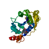
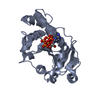
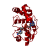
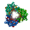
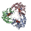
| Title | Crystal Structure of Nicotinic Acid Mononucleotide Adenylyltransferase from Pseudomonas aeruginosa | ||||||
 Components Components | 'Probable nicotinate-nucleotide adenylyltransferase | ||||||
 Keywords Keywords | TRANSFERASE / alpha/beta domain | ||||||
| Function / homology |  Function and homology information Function and homology informationnicotinate-nucleotide adenylyltransferase / nicotinamide-nucleotide adenylyltransferase activity / nicotinate-nucleotide adenylyltransferase activity / NAD+ biosynthetic process via the salvage pathway / ATP binding Similarity search - Function | ||||||
| Biological species |   Pseudomonas aeruginosa (bacteria) Pseudomonas aeruginosa (bacteria) | ||||||
| Method |  X-RAY DIFFRACTION / SYNCHROTRON / MOLECULAR REPLACEMENT / Resolution: 1.7 Å X-RAY DIFFRACTION / SYNCHROTRON / MOLECULAR REPLACEMENT / Resolution: 1.7 Å | ||||||
 Authors Authors | Yoon, H.J. / Kim, H.L. / Mikami, B. / Suh, S.W. | ||||||
 Citation Citation | Journal: J.Mol.Biol. / Year: 2005 Title: Crystal structure of nicotinic acid mononucleotide adenylyltransferase from Pseudomonas aeruginosa in its Apo and substrate-complexed forms reveals a fully open conformation Authors: Yoon, H.J. / Kim, H.L. / Mikami, B. / Suh, S.W. | ||||||
| History |
| ||||||
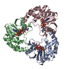
| Remark 300 | BIOMOLECULE: 1 THIS ENTRY CONTAINS THE CRYSTALLOGRAPHIC ASYMMETRIC UNIT WHICH CONSISTS OF 4 CHAINS. ...BIOMOLECULE: 1 THIS ENTRY CONTAINS THE CRYSTALLOGRAPHIC ASYMMETRIC UNIT WHICH CONSISTS OF 4 CHAINS. THE BIOLOGICAL UNIT IS NOT ASSIGNED. |
- Structure visualization
| Structure viewer | Molecule: MolmilJmol/JSmol |
|---|
- Downloads & links
-Download
| PDBx/mmCIF format | 1yum.cif.gz | 204.2 KB | Display | PDBx/mmCIF format |
|---|---|---|---|---|
| PDB format | pdb1yum.ent.gz | 161.8 KB | Display | PDB format |
| PDBx/mmJSON format | 1yum.json.gz | Tree view | PDBx/mmJSON format | |
| Others |  Other downloads Other downloads |
-Validation report
| Arichive directory | https://data.pdbj.org/pub/pdb/validation_reports/yu/1yumftp://data.pdbj.org/pub/pdb/validation_reports/yu/1yum | HTTPS FTP |
|---|
-Related structure data


-Links
 PDBj
PDBj- Assembly
| Deposited unit | 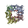
| ||||||||
|---|---|---|---|---|---|---|---|---|---|
| 1 |
| ||||||||
| Unit cell |
| ||||||||
| Details | The biological assembly is a dimer generated from the tetramer in the asymmetric unit by the operations: -x, 1/2+y, -z |
-Components
| #1: Protein | ' Mass: 27074.783 Da / Num. of mol.: 4 Source method: isolated from a genetically manipulated source Source: (gene. exp.) Pseudomonas aeruginosa (bacteria) / Gene: nadD (PA4006) / Plasmid: pET-28b(+) / Production host: References: UniProt: Q9HX21, nicotinate-nucleotide adenylyltransferase #2: Chemical | ChemComp-CIT /   Mass: 192.124 Da / Num. of mol.: 4 / Source method: obtained synthetically / Formula: C6H8O7 Mass: 192.124 Da / Num. of mol.: 4 / Source method: obtained synthetically / Formula: C6H8O7#3: Chemical | ChemComp-NCN /   Mass: 335.204 Da / Num. of mol.: 4 / Source method: obtained synthetically / Formula: C11H14NO9P Mass: 335.204 Da / Num. of mol.: 4 / Source method: obtained synthetically / Formula: C11H14NO9P#4: Water | ChemComp-HOH / |  Mass: 18.015 Da / Num. of mol.: 813 / Source method: isolated from a natural source / Formula: H2O Mass: 18.015 Da / Num. of mol.: 813 / Source method: isolated from a natural source / Formula: H2O |
|---|
-Experimental details
-Experiment
| Experiment | Method: X-RAY DIFFRACTION / Number of used crystals: 1 |
|---|
- Sample preparation
| Crystal | Density Matthews: 2.15 Å3/Da / Density % sol: 42.7 % |
|---|---|
| Crystal grow | Temperature: 291 K / Method: vapor diffusion, hanging drop / pH: 7 Details: bis-tris propane, trisodium citrate, glycerol, pH 7.0, VAPOR DIFFUSION, HANGING DROP, temperature 291K |
-Data collection
| Diffraction | Mean temperature: 100 K |
|---|---|
| Diffraction source | Source: SYNCHROTRON / Site: SPring-8  / Beamline: BL38B1 / Wavelength: 0.85 Å / Beamline: BL38B1 / Wavelength: 0.85 Å |
| Detector | Type: ADSC QUANTUM 4 / Detector: CCD / Date: Feb 16, 2004 / Details: mirrors |
| Radiation | Monochromator: quasi-monochromatic X-ray / Protocol: SINGLE WAVELENGTH / Monochromatic (M) / Laue (L): M / Scattering type: x-ray |
| Radiation wavelength | Wavelength: 0.85 Å / Relative weight: 1 |
| Reflection | Resolution: 1.6→50 Å / Num. all: 118067 / Num. obs: 118041 / % possible obs: 96.8 % / Observed criterion σ(I): -3 / Redundancy: 3.1 % / Biso Wilson estimate: 21.74 Å2 / Rmerge(I) obs: 0.1 / Net I/σ(I): 23.5 |
| Reflection shell | Resolution: 1.6→1.66 Å / Redundancy: 3 % / Rmerge(I) obs: 0.266 / Num. unique all: 12046 / % possible all: 98.9 |
- Processing
| Software |
| |||||||||||||||||||||||||||||||||||
|---|---|---|---|---|---|---|---|---|---|---|---|---|---|---|---|---|---|---|---|---|---|---|---|---|---|---|---|---|---|---|---|---|---|---|---|---|
| Refinement | Method to determine structure: MOLECULAR REPLACEMENT Starting model: apo NaMN AT Resolution: 1.7→10 Å / Num. parameters: 31002 / Num. restraintsaints: 29215 / Cross valid method: THROUGHOUT / σ(I): 0 Details: THIS IS A TWINNED STRUCTURE. THE TWINNING OPERATOR IS (H,K,L) -> (H,-K,-L) AND THE TWINNING FRACTION IS 0.49421
| |||||||||||||||||||||||||||||||||||
| Refine analyze | Num. disordered residues: 26 / Occupancy sum hydrogen: 0 / Occupancy sum non hydrogen: 7590.14 | |||||||||||||||||||||||||||||||||||
| Refinement step | Cycle: LAST / Resolution: 1.7→10 Å
| |||||||||||||||||||||||||||||||||||
| Refine LS restraints |
| |||||||||||||||||||||||||||||||||||
| LS refinement shell |
|
