 ムービー
ムービー コントローラー


+ データを開く

- 基本情報
基本情報
| 登録情報 | データベース: PDB / ID: 1hjt | ||||||
|---|---|---|---|---|---|---|---|
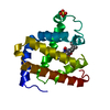
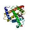
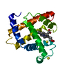
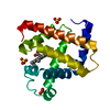
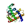
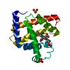
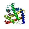
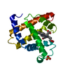
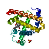
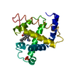
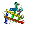
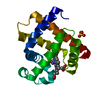
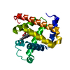
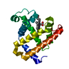
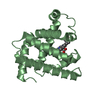
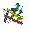
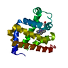
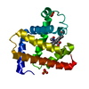
| タイトル | SPERM WHALE MYOGLOBIN (FERROUS, NITRIC OXIDE BOUND) | ||||||
 要素 要素 | MYOGLOBIN | ||||||
 キーワード キーワード | OXYGEN TRANSPORT / GLOBIN / HEME / OXYGEN STORAGE / NITRIC OXIDE | ||||||
| 機能・相同性 |  機能・相同性情報 機能・相同性情報酸化還元酵素; 他の含窒素化合物が電子供与する / nitrite reductase activity / sarcoplasm / 酸化還元酵素; 過酸化物を電子受容体にする; ペルオキシダーゼ / removal of superoxide radicals / oxygen carrier activity / peroxidase activity / oxygen binding / heme binding / extracellular exosome / metal ion binding 類似検索 - 分子機能 | ||||||
| 生物種 |  | ||||||
| 手法 |  X線回折 / DIFFERENCE FOURIER / 解像度: 1.7 Å X線回折 / DIFFERENCE FOURIER / 解像度: 1.7 Å | ||||||
 データ登録者 データ登録者 | Brucker, E.A. / Phillips Jr., G.N. | ||||||
 引用 引用 | ジャーナル: Proteins / 年: 1998 タイトル: Nitric oxide myoglobin: crystal structure and analysis of ligand geometry. 著者: Brucker, E.A. / Olson, J.S. / Ikeda-Saito, M. / Phillips Jr., G.N. | ||||||
| 履歴 |
|
- 構造の表示
構造の表示
| 構造ビューア | 分子: MolmilJmol/JSmol |
|---|
- ダウンロードとリンク
-ダウンロード
| PDBx/mmCIF形式 | 1hjt.cif.gz | 46.4 KB | 表示 | PDBx/mmCIF形式 |
|---|---|---|---|---|
| PDB形式 | pdb1hjt.ent.gz | 32 KB | 表示 | PDB形式 |
| PDBx/mmJSON形式 | 1hjt.json.gz | ツリー表示 | PDBx/mmJSON形式 | |
| その他 |  その他のダウンロード その他のダウンロード |
-検証レポート
| アーカイブディレクトリ | https://data.pdbj.org/pub/pdb/validation_reports/hj/1hjtftp://data.pdbj.org/pub/pdb/validation_reports/hj/1hjt | HTTPS FTP |
|---|
-関連構造データ


-リンク
 PDBj
PDBj


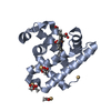
- 集合体
集合体
| 登録構造単位 | 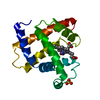
| ||||||||
|---|---|---|---|---|---|---|---|---|---|
| 1 |
| ||||||||
| 単位格子 |
|
-要素
| #1: タンパク質 | 分子量: 17234.951 Da / 分子数: 1 / 由来タイプ: 天然 / 由来: (天然) |
|---|---|
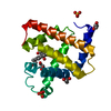
| #2: 化合物 | ChemComp-SO4 /   分子量: 96.063 Da / 分子数: 1 / 由来タイプ: 合成 / 式: SO4 分子量: 96.063 Da / 分子数: 1 / 由来タイプ: 合成 / 式: SO4 |
| #3: 化合物 | ChemComp-HEM /   分子量: 616.487 Da / 分子数: 1 / 由来タイプ: 合成 / 式: C34H32FeN4O4 分子量: 616.487 Da / 分子数: 1 / 由来タイプ: 合成 / 式: C34H32FeN4O4 |
| #4: 化合物 | ChemComp-NO /   分子量: 30.006 Da / 分子数: 1 / 由来タイプ: 合成 / 式: NO 分子量: 30.006 Da / 分子数: 1 / 由来タイプ: 合成 / 式: NO |
| #5: 水 | ChemComp-HOH /  分子量: 18.015 Da / 分子数: 106 / 由来タイプ: 天然 / 式: H2O 分子量: 18.015 Da / 分子数: 106 / 由来タイプ: 天然 / 式: H2O |
-実験情報
-実験
| 実験 | 手法: X線回折 / 使用した結晶の数: 1 |
|---|
- 試料調製
試料調製
| 結晶 | マシュー密度: 1.87 Å3/Da / 溶媒含有率: 34.09 % | |||||||||||||||||||||||||
|---|---|---|---|---|---|---|---|---|---|---|---|---|---|---|---|---|---|---|---|---|---|---|---|---|---|---|
| 結晶化 | *PLUS 温度: 17 ℃ / 手法: batch method | |||||||||||||||||||||||||
| 溶液の組成 | *PLUS
|
-データ収集
| 回折 | 平均測定温度: 295 K |
|---|---|
| 放射光源 | 由来: 回転陽極 / タイプ: SIEMENS / 波長: 1.5418 |
| 検出器 | タイプ: RIGAKU / 検出器: IMAGE PLATE / 日付: 1997年3月10日 |
| 放射 | 単色(M)・ラウエ(L): M / 散乱光タイプ: x-ray |
| 放射波長 | 波長: 1.5418 Å / 相対比: 1 |
| 反射 | 解像度: 1.7→30 Å / Num. obs: 11674 / % possible obs: 80.1 % / 冗長度: 3.1 % / Rmerge(I) obs: 0.044 / Net I/σ(I): 32.7 |
| 反射 シェル | 解像度: 1.7→1.8 Å / 冗長度: 2.5 % / Rmerge(I) obs: 0.244 / Mean I/σ(I) obs: 6.4 / % possible all: 73.1 |
| 反射 | *PLUS Num. measured all: 56610 |
| 反射 シェル | *PLUS % possible obs: 73.1 % |
- 解析
解析
| ソフトウェア |
| |||||||||||||||||||||||||||||||||
|---|---|---|---|---|---|---|---|---|---|---|---|---|---|---|---|---|---|---|---|---|---|---|---|---|---|---|---|---|---|---|---|---|---|---|
| 精密化 | 構造決定の手法: DIFFERENCE FOURIER 開始モデル: PDB ENTRY 1YOI 解像度: 1.7→5 Å / Num. parameters: 5499 / Num. restraintsaints: 5369 / 交差検証法: FREE R / 立体化学のターゲット値: ENGH AND HUBER
| |||||||||||||||||||||||||||||||||
| 溶媒の処理 | 溶媒モデル: MOEWS & KRETSINGER, J.MOL.BIOL.91(1973)201-228 | |||||||||||||||||||||||||||||||||
| Refine analyze | Num. disordered residues: 0 / Occupancy sum hydrogen: 0 / Occupancy sum non hydrogen: 1352.4 | |||||||||||||||||||||||||||||||||
| 精密化ステップ | サイクル: LAST / 解像度: 1.7→5 Å
| |||||||||||||||||||||||||||||||||
| 拘束条件 |
| |||||||||||||||||||||||||||||||||
| ソフトウェア | *PLUS 名称: SHELXL-96 / 分類: refinement | |||||||||||||||||||||||||||||||||
| 拘束条件 | *PLUS
|
