 Movie
Movie Controller

[English] 日本語
 Yorodumi
- PDB-1hdq: Crystal structure of bovine pancreatic carboxypeptidase A complex... -
+ Open data

- Basic information
| Entry | Database: PDB / ID: 1hdq | ||||||
|---|---|---|---|---|---|---|---|
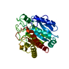
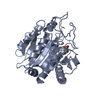
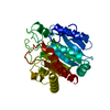
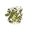
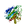
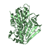
| Title | Crystal structure of bovine pancreatic carboxypeptidase A complexed with D-N-hydroxyaminocarbonyl phenylalanine at 2.3 A | ||||||
 Components Components | CARBOXYPEPTIDASE A | ||||||
 Keywords Keywords | CARBOXYPEPTIDASE / CPA / LBHB / INHIBITOR | ||||||
| Function / homology |  Function and homology informationcarboxypeptidase A / leukotriene metabolic process / metallocarboxypeptidase activity / proteolysis / extracellular space / zinc ion binding Function and homology informationcarboxypeptidase A / leukotriene metabolic process / metallocarboxypeptidase activity / proteolysis / extracellular space / zinc ion bindingSimilarity search - Function | ||||||
| Biological species |  BOS BOVIS (cattle) BOS BOVIS (cattle) | ||||||
| Method | X-RAY DIFFRACTION / OTHER / Resolution: 2.3 Å | ||||||
 Authors Authors | Cho, J.H. / Ha, N.-C. / Chung, S.J. / Kim, D.H. / Choi, K.Y. / Oh, B.-H. | ||||||
 Citation Citation | Journal: Bioorg.Med.Chem. / Year: 2002 Title: Insight Into the Stereochemistry in the Inhibition of Carboxypeptidase a with N-(Hydroxyaminocarbonyl)Phenylalanine: Binding Modes of an Enantiomeric Pair of the Inhibitor to Carboxypeptidase A Authors: Cho, J.H. / Kim, D.H. / Chung, S.J. / Ha, N.-C. / Oh, B.-H. / Choi, K.Y. | ||||||
| History |
|
- Structure visualization
| Structure viewer | Molecule: MolmilJmol/JSmol |
|---|
- Downloads & links
-Download
| PDBx/mmCIF format | 1hdq.cif.gz | 74.2 KB | Display | PDBx/mmCIF format |
|---|---|---|---|---|
| PDB format | pdb1hdq.ent.gz | 58.5 KB | Display | PDB format |
| PDBx/mmJSON format | 1hdq.json.gz | Tree view | PDBx/mmJSON format | |
| Others |  Other downloads Other downloads |
-Validation report
| Arichive directory | https://data.pdbj.org/pub/pdb/validation_reports/hd/1hdqftp://data.pdbj.org/pub/pdb/validation_reports/hd/1hdq | HTTPS FTP |
|---|
-Related structure data


-Links
 PDBj
PDBj
- Assembly
| Deposited unit | 
| ||||||||
|---|---|---|---|---|---|---|---|---|---|
| 1 |
| ||||||||
| Unit cell |
| ||||||||
| Details | BIOLOGICAL_UNIT: MONOMER |
-Components
| #1: Protein | Mass: 34517.480 Da / Num. of mol.: 1 / Source method: isolated from a natural source / Source: (natural) BOS BOVIS (cattle) / Organ: PANCREAS / References: UniProt: P00730, carboxypeptidase A |
|---|---|
| #2: Chemical | ChemComp-INF /   Mass: 224.213 Da / Num. of mol.: 1 / Source method: obtained synthetically / Formula: C10H12N2O4 Mass: 224.213 Da / Num. of mol.: 1 / Source method: obtained synthetically / Formula: C10H12N2O4 |
| #3: Chemical | ChemComp-ZN /   Mass: 65.409 Da / Num. of mol.: 1 / Source method: obtained synthetically / Formula: Zn Mass: 65.409 Da / Num. of mol.: 1 / Source method: obtained synthetically / Formula: Zn |
| #4: Water | ChemComp-HOH / Water Mass: 18.015 Da / Num. of mol.: 211 / Source method: isolated from a natural source / Formula: H2O Mass: 18.015 Da / Num. of mol.: 211 / Source method: isolated from a natural source / Formula: H2O |
| Compound details | PEPTIDYL-L-AMINO ACID + H(2)O = PEPTIDE + L-AMINO ACID THE ZYMOGEN IS SECRETED AS A TERNARY COMPLEX ...PEPTIDYL-L-AMINO ACID + H(2)O = PEPTIDE + L-AMINO ACID THE ZYMOGEN IS SECRETED AS A TERNARY COMPLEX COMPOSED OF PROCARBOXY |
-Experimental details
-Experiment
| Experiment | Method: X-RAY DIFFRACTION / Number of used crystals: 1 |
|---|
- Sample preparation
| Crystal | Density Matthews: 2.16 Å3/Da / Density % sol: 42.95 % | |||||||||||||||||||||||||||||||||||
|---|---|---|---|---|---|---|---|---|---|---|---|---|---|---|---|---|---|---|---|---|---|---|---|---|---|---|---|---|---|---|---|---|---|---|---|---|
| Crystal grow | pH: 7.5 / Details: pH 7.50 | |||||||||||||||||||||||||||||||||||
| Crystal grow | *PLUS Method: microdialysis | |||||||||||||||||||||||||||||||||||
| Components of the solutions | *PLUS
|
-Data collection
| Diffraction | Mean temperature: 297 K |
|---|---|
| Diffraction source | Source: ROTATING ANODE / Wavelength: 1.5418 |
| Radiation | Protocol: SINGLE WAVELENGTH / Monochromatic (M) / Laue (L): M / Scattering type: x-ray |
| Radiation wavelength | Wavelength: 1.5418 Å / Relative weight: 1 |
| Reflection | Resolution: 2.3→20 Å / Num. obs: 13199 / % possible obs: 96 % / Observed criterion σ(I): 2 / Redundancy: 2.5 % / Rmerge(I) obs: 0.06 / Rsym value: 0.06 / Net I/σ(I): 33 |
| Reflection | *PLUS Highest resolution: 2.3 Å / Lowest resolution: 20 Å / % possible obs: 96 % / Rmerge(I) obs: 0.06 |
| Reflection shell | *PLUS Highest resolution: 2.3 Å / Lowest resolution: 2.38 Å / % possible obs: 82.2 % / Rmerge(I) obs: 0.094 |
- Processing
| Software | Name: CNS / Version: 1 / Classification: refinement | ||||||||||||||||||||||||||||||||||||||||||||||||||||||||||||
|---|---|---|---|---|---|---|---|---|---|---|---|---|---|---|---|---|---|---|---|---|---|---|---|---|---|---|---|---|---|---|---|---|---|---|---|---|---|---|---|---|---|---|---|---|---|---|---|---|---|---|---|---|---|---|---|---|---|---|---|---|---|
| Refinement | Method to determine structure: OTHER / Resolution: 2.3→100 Å / Cross valid method: THROUGHOUT / σ(F): 0
| ||||||||||||||||||||||||||||||||||||||||||||||||||||||||||||
| Refinement step | Cycle: LAST / Resolution: 2.3→100 Å
| ||||||||||||||||||||||||||||||||||||||||||||||||||||||||||||
| Refine LS restraints |
| ||||||||||||||||||||||||||||||||||||||||||||||||||||||||||||
| Software | *PLUS Version: 1 / Classification: refinement | ||||||||||||||||||||||||||||||||||||||||||||||||||||||||||||
| Refinement | *PLUS Lowest resolution: 100 Å | ||||||||||||||||||||||||||||||||||||||||||||||||||||||||||||
| Solvent computation | *PLUS | ||||||||||||||||||||||||||||||||||||||||||||||||||||||||||||
| Displacement parameters | *PLUS | ||||||||||||||||||||||||||||||||||||||||||||||||||||||||||||
| Refine LS restraints | *PLUS Type: c_angle_deg / Dev ideal: 1.172 |
