 Movie
Movie Controller

[English] 日本語
 Yorodumi
- PDB-2ctc: THE HIGH RESOLUTION CRYSTAL STRUCTURE OF THE COMPLEX BETWEEN CARB... -
+ Open data

- Basic information
| Entry | Database: PDB / ID: 2ctc | ||||||
|---|---|---|---|---|---|---|---|
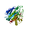
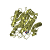
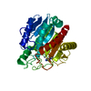
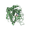
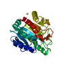
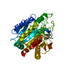
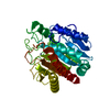
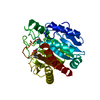
| Title | THE HIGH RESOLUTION CRYSTAL STRUCTURE OF THE COMPLEX BETWEEN CARBOXYPEPTIDASE A AND L-PHENYL LACTATE | ||||||
 Components Components | CARBOXYPEPTIDASE A | ||||||
 Keywords Keywords | HYDROLASE(C-TERMINAL PEPTIDASE) | ||||||
| Function / homology |  Function and homology information Function and homology informationcarboxypeptidase A / leukotriene metabolic process / metallocarboxypeptidase activity / proteolysis / : / zinc ion binding Similarity search - Function | ||||||
| Biological species |  | ||||||
| Method |  X-RAY DIFFRACTION / Resolution: 1.4 Å X-RAY DIFFRACTION / Resolution: 1.4 Å | ||||||
 Authors Authors | Teplyakov, A. / Wilson, K.S. / Orioli, P. / Mangani, S. | ||||||
 Citation Citation | Journal: Acta Crystallogr.,Sect.D / Year: 1993 Title: High-resolution structure of the complex between carboxypeptidase A and L-phenyl lactate. Authors: Teplyakov, A. / Wilson, K.S. / Orioli, P. / Mangani, S. | ||||||
| History |
|
- Structure visualization
| Structure viewer | Molecule: MolmilJmol/JSmol |
|---|
- Downloads & links
-Download
| PDBx/mmCIF format | 2ctc.cif.gz | 76.7 KB | Display | PDBx/mmCIF format |
|---|---|---|---|---|
| PDB format | pdb2ctc.ent.gz | 56.6 KB | Display | PDB format |
| PDBx/mmJSON format | 2ctc.json.gz | Tree view | PDBx/mmJSON format | |
| Others |  Other downloads Other downloads |
-Validation report
| Arichive directory | https://data.pdbj.org/pub/pdb/validation_reports/ct/2ctcftp://data.pdbj.org/pub/pdb/validation_reports/ct/2ctc | HTTPS FTP |
|---|
-Related structure data


-Links
 PDBj
PDBj
- Assembly
| Deposited unit | 
| ||||||||
|---|---|---|---|---|---|---|---|---|---|
| 1 |
| ||||||||
| Unit cell |
| ||||||||
| Atom site foot note | 1: PEPTIDE BONDS BETWEEN RESIDUES SER 197 - TYR 198, PRO 205 - TYR 206, AND ARG 272 - ASP 273 ARE IN CIS CONFORMATION. |
-Components
| #1: Protein | Mass: 34517.480 Da / Num. of mol.: 1 Source method: isolated from a genetically manipulated source Source: (gene. exp.) |
|---|---|
| #2: Chemical | ChemComp-ZN /   Mass: 65.409 Da / Num. of mol.: 1 / Source method: obtained synthetically / Formula: Zn Mass: 65.409 Da / Num. of mol.: 1 / Source method: obtained synthetically / Formula: Zn |
| #3: Chemical | ChemComp-HFA /   Type: L-peptide linking / Mass: 166.174 Da / Num. of mol.: 1 / Source method: obtained synthetically / Formula: C9H10O3 Type: L-peptide linking / Mass: 166.174 Da / Num. of mol.: 1 / Source method: obtained synthetically / Formula: C9H10O3 |
| #4: Water | ChemComp-HOH /  Mass: 18.015 Da / Num. of mol.: 181 / Source method: isolated from a natural source / Formula: H2O Mass: 18.015 Da / Num. of mol.: 181 / Source method: isolated from a natural source / Formula: H2O |
| Has protein modification | Y |
-Experimental details
-Experiment
| Experiment | Method: X-RAY DIFFRACTION |
|---|
- Sample preparation
| Crystal | Density Matthews: 2.11 Å3/Da / Density % sol: 41.73 % | ||||||||||||||||||
|---|---|---|---|---|---|---|---|---|---|---|---|---|---|---|---|---|---|---|---|
| Crystal grow | *PLUS pH: 7.5 / Method: vapor diffusion, hanging drop / Details: referred to J.Mol.Biol. 19.423-441 1966 | ||||||||||||||||||
| Components of the solutions | *PLUS
|
-Data collection
| Reflection | *PLUS Highest resolution: 1.45 Å / Lowest resolution: 3.5 Å / Num. obs: 46136 / % possible obs: 90.4 % / Num. measured all: 127939 / Rmerge(I) obs: 0.036 |
|---|
- Processing
| Software | Name: PROLSQ / Classification: refinement | ||||||||||||||||||
|---|---|---|---|---|---|---|---|---|---|---|---|---|---|---|---|---|---|---|---|
| Refinement | Highest resolution: 1.4 Å /
| ||||||||||||||||||
| Refinement step | Cycle: LAST / Highest resolution: 1.4 Å
| ||||||||||||||||||
| Software | *PLUS Name: PROLSQ / Classification: refinement | ||||||||||||||||||
| Refinement | *PLUS Highest resolution: 1.45 Å / Num. reflection obs: 45829 / Rfactor obs: 0.161 / Lowest resolution: 8 Å | ||||||||||||||||||
| Solvent computation | *PLUS | ||||||||||||||||||
| Displacement parameters | *PLUS | ||||||||||||||||||
| Refine LS restraints | *PLUS
|
