 Movie
Movie Controller

[English] 日本語
 Yorodumi
- PDB-1h9u: The structure of the human retinoid-X-receptor beta ligand bindin... -
+ Open data

- Basic information
| Entry | Database: PDB / ID: 1h9u | ||||||
|---|---|---|---|---|---|---|---|
| Title | The structure of the human retinoid-X-receptor beta ligand binding domain in complex with the specific synthetic agonist LG100268 | ||||||
 Components Components | RETINOID X RECEPTOR, BETA | ||||||
 Keywords Keywords | NUCLEAR RECEPTOR / RXR / TRANSCRIPTION FACTOR | ||||||
| Function / homology |  Function and homology information Function and homology informationventricular cardiac muscle cell differentiation / maternal placenta development / retinoic acid-responsive element binding / NR1H2 & NR1H3 regulate gene expression linked to triglyceride lipolysis in adipose / NR1H2 & NR1H3 regulate gene expression linked to gluconeogenesis / NR1H2 & NR1H3 regulate gene expression to limit cholesterol uptake / NR1H2 & NR1H3 regulate gene expression linked to lipogenesis / positive regulation of vitamin D receptor signaling pathway / Signaling by Retinoic Acid / NR1H2 & NR1H3 regulate gene expression to control bile acid homeostasis ...ventricular cardiac muscle cell differentiation / maternal placenta development / retinoic acid-responsive element binding / NR1H2 & NR1H3 regulate gene expression linked to triglyceride lipolysis in adipose / NR1H2 & NR1H3 regulate gene expression linked to gluconeogenesis / NR1H2 & NR1H3 regulate gene expression to limit cholesterol uptake / NR1H2 & NR1H3 regulate gene expression linked to lipogenesis / positive regulation of vitamin D receptor signaling pathway / Signaling by Retinoic Acid / NR1H2 & NR1H3 regulate gene expression to control bile acid homeostasis / nuclear steroid receptor activity / cardiac muscle cell proliferation / positive regulation of bone mineralization / retinoic acid receptor signaling pathway / NR1H3 & NR1H2 regulate gene expression linked to cholesterol transport and efflux / cellular response to retinoic acid / hormone-mediated signaling pathway / RNA polymerase II transcription regulatory region sequence-specific DNA binding / PPARA activates gene expression / chromatin DNA binding / Nuclear Receptor transcription pathway / mRNA transcription by RNA polymerase II / RNA polymerase II transcription regulator complex / nuclear receptor activity / sequence-specific double-stranded DNA binding / nervous system development / DNA-binding transcription activator activity, RNA polymerase II-specific / in utero embryonic development / DNA-binding transcription factor activity, RNA polymerase II-specific / cell differentiation / positive regulation of DNA-templated transcription / chromatin / nucleolus / positive regulation of transcription by RNA polymerase II / zinc ion binding / nucleoplasm / nucleus / cytosol Similarity search - Function | ||||||
| Biological species |  HOMO SAPIENS (human) HOMO SAPIENS (human) | ||||||
| Method |  X-RAY DIFFRACTION / SYNCHROTRON / MOLECULAR REPLACEMENT / Resolution: 2.7 Å X-RAY DIFFRACTION / SYNCHROTRON / MOLECULAR REPLACEMENT / Resolution: 2.7 Å | ||||||
 Authors Authors | Schwabe, J.W.R. / Love, J.D. / Gooch, J.T. | ||||||
 Citation Citation | Journal: J.Biol.Chem. / Year: 2002 Title: The Structural Basis for the Specificity of Retinoid-X Receptor-Selective Agonists: New Insights Into the Role of Helix H12 Authors: Love, J.D. / Gooch, J.T. / Benko, S. / Li, C. / Nagy, L. / Chatterjee, V.K.K. / Evans, R.M. / Schwabe, J.W.R. | ||||||
| History |
|
- Structure visualization
| Structure viewer | Molecule: MolmilJmol/JSmol |
|---|
- Downloads & links
-Download
| PDBx/mmCIF format | 1h9u.cif.gz | 167.9 KB | Display | PDBx/mmCIF format |
|---|---|---|---|---|
| PDB format | pdb1h9u.ent.gz | 132.7 KB | Display | PDB format |
| PDBx/mmJSON format | 1h9u.json.gz | Tree view | PDBx/mmJSON format | |
| Others |  Other downloads Other downloads |
-Validation report
| Arichive directory | https://data.pdbj.org/pub/pdb/validation_reports/h9/1h9uftp://data.pdbj.org/pub/pdb/validation_reports/h9/1h9u | HTTPS FTP |
|---|
-Related structure data
| Related structure data |  1lbd S: Starting model for refinement |
|---|---|
| Similar structure data |


-Links
 PDBj
PDBj


- Assembly
| Deposited unit | 
| ||||||||
|---|---|---|---|---|---|---|---|---|---|
| 1 | 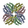
| ||||||||
| Unit cell |
| ||||||||
| Details | CRYSTAL. THE RETINOID X RECEPTOR IS ACTIVE AS DIMERIC |
-Components
| #1: Protein | Mass: 24842.645 Da / Num. of mol.: 4 / Fragment: LIGAND BINDING DOMAIN RESIDUES 299-522 Source method: isolated from a genetically manipulated source Source: (gene. exp.) HOMO SAPIENS (human) / Production host:  #2: Chemical | ChemComp-LG2 /   Mass: 363.493 Da / Num. of mol.: 4 / Source method: obtained synthetically / Formula: C24H29NO2 Mass: 363.493 Da / Num. of mol.: 4 / Source method: obtained synthetically / Formula: C24H29NO2#3: Chemical | ChemComp-NI /   Mass: 58.693 Da / Num. of mol.: 4 / Source method: obtained synthetically / Formula: Ni Mass: 58.693 Da / Num. of mol.: 4 / Source method: obtained synthetically / Formula: Ni#4: Chemical |   Mass: 35.453 Da / Num. of mol.: 2 / Source method: obtained synthetically / Formula: Cl Mass: 35.453 Da / Num. of mol.: 2 / Source method: obtained synthetically / Formula: Cl#5: Water | ChemComp-HOH / |  Mass: 18.015 Da / Num. of mol.: 4 / Source method: isolated from a natural source / Formula: H2O Mass: 18.015 Da / Num. of mol.: 4 / Source method: isolated from a natural source / Formula: H2OCompound details | BINDS TO 9-CIS RETINOIC ACID (9C-RA). INVOLVED IN RETINOIC ACID RESPONSE PATHWAY. THE C-TERMINAL ...BINDS TO 9-CIS RETINOIC ACID (9C-RA). INVOLVED IN RETINOIC ACID RESPONSE PATHWAY. THE C-TERMINAL DOMAIN IS INVOLVED IN STEROID-BINDING. | |
|---|
-Experimental details
-Experiment
| Experiment | Method: X-RAY DIFFRACTION / Number of used crystals: 1 |
|---|
- Sample preparation
| Crystal | Density Matthews: 2.82 Å3/Da / Density % sol: 56.45 % | |||||||||||||||||||||||||||||||||||||||||||||||||
|---|---|---|---|---|---|---|---|---|---|---|---|---|---|---|---|---|---|---|---|---|---|---|---|---|---|---|---|---|---|---|---|---|---|---|---|---|---|---|---|---|---|---|---|---|---|---|---|---|---|---|
| Crystal grow | pH: 7.6 / Details: pH 7.60 | |||||||||||||||||||||||||||||||||||||||||||||||||
| Crystal grow | *PLUS Temperature: 20 ℃ / Method: vapor diffusion | |||||||||||||||||||||||||||||||||||||||||||||||||
| Components of the solutions | *PLUS
|
-Data collection
| Diffraction | Mean temperature: 100 K |
|---|---|
| Diffraction source | Source: SYNCHROTRON / Site: ESRF  / Beamline: ID14-2 / Wavelength: 0.933 / Beamline: ID14-2 / Wavelength: 0.933 |
| Detector | Type: QUANTUM / Detector: CCD |
| Radiation | Protocol: SINGLE WAVELENGTH / Monochromatic (M) / Laue (L): M / Scattering type: x-ray |
| Radiation wavelength | Wavelength: 0.933 Å / Relative weight: 1 |
| Reflection | Resolution: 2.7→38 Å / Num. obs: 29713 / % possible obs: 98 % / Observed criterion σ(I): 2 / Redundancy: 2.9 % / Biso Wilson estimate: 55.3 Å2 / Rmerge(I) obs: 0.098 / Net I/σ(I): 5.1 |
| Reflection | *PLUS Lowest resolution: 38 Å / % possible obs: 97.8 % / Num. measured all: 87647 |
| Reflection shell | *PLUS Highest resolution: 2.7 Å / Lowest resolution: 2.87 Å / Rmerge(I) obs: 0.264 / Mean I/σ(I) obs: 2.1 |
- Processing
| Software |
| ||||||||||||||||||||||||||||||||||||||||||||||||||||||||||||||||||||||||||||||||
|---|---|---|---|---|---|---|---|---|---|---|---|---|---|---|---|---|---|---|---|---|---|---|---|---|---|---|---|---|---|---|---|---|---|---|---|---|---|---|---|---|---|---|---|---|---|---|---|---|---|---|---|---|---|---|---|---|---|---|---|---|---|---|---|---|---|---|---|---|---|---|---|---|---|---|---|---|---|---|---|---|---|
| Refinement | Method to determine structure: MOLECULAR REPLACEMENT Starting model: PDB ENTRY 1LBD 1lbd Resolution: 2.7→38.14 Å / Rfactor Rfree error: 0.006 / Data cutoff high absF: 2064073.76 / Isotropic thermal model: RESTRAINED / Cross valid method: THROUGHOUT / σ(F): 2
| ||||||||||||||||||||||||||||||||||||||||||||||||||||||||||||||||||||||||||||||||
| Solvent computation | Solvent model: FLAT MODEL / Bsol: 52.2394 Å2 / ksol: 0.35528 e/Å3 | ||||||||||||||||||||||||||||||||||||||||||||||||||||||||||||||||||||||||||||||||
| Displacement parameters | Biso mean: 50.5 Å2
| ||||||||||||||||||||||||||||||||||||||||||||||||||||||||||||||||||||||||||||||||
| Refine analyze |
| ||||||||||||||||||||||||||||||||||||||||||||||||||||||||||||||||||||||||||||||||
| Refinement step | Cycle: LAST / Resolution: 2.7→38.14 Å
| ||||||||||||||||||||||||||||||||||||||||||||||||||||||||||||||||||||||||||||||||
| Refine LS restraints |
| ||||||||||||||||||||||||||||||||||||||||||||||||||||||||||||||||||||||||||||||||
| LS refinement shell | Resolution: 2.7→2.87 Å / Rfactor Rfree error: 0.018 / Total num. of bins used: 6
| ||||||||||||||||||||||||||||||||||||||||||||||||||||||||||||||||||||||||||||||||
| Xplor file |
| ||||||||||||||||||||||||||||||||||||||||||||||||||||||||||||||||||||||||||||||||
| Refinement | *PLUS Rfactor obs: 0.277 / Rfactor Rfree: 0.301 / Rfactor Rwork: 0.277 | ||||||||||||||||||||||||||||||||||||||||||||||||||||||||||||||||||||||||||||||||
| Solvent computation | *PLUS | ||||||||||||||||||||||||||||||||||||||||||||||||||||||||||||||||||||||||||||||||
| Displacement parameters | *PLUS | ||||||||||||||||||||||||||||||||||||||||||||||||||||||||||||||||||||||||||||||||
| Refine LS restraints | *PLUS
|
