 Movie
Movie Controller

[English] 日本語
 Yorodumi
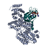
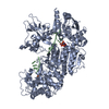
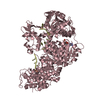
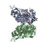
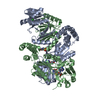
- PDB-1h2t: Structure of the human nuclear cap-binding-complex (CBC) in compl... -
+ Open data

- Basic information
| Entry | Database: PDB / ID: 1h2t | ||||||
|---|---|---|---|---|---|---|---|
| Title | Structure of the human nuclear cap-binding-complex (CBC) in complex with a cap analogue m7GpppG | ||||||
 Components Components |
| ||||||
 Keywords Keywords | NUCLEAR PROTEIN / M7GCAP / CAP-BINDING-COMPLEX / RNP DOMAIN / MIF4G DOMAIN / RNA MATURATION / RNA EXPORT / RNA-BINDING | ||||||
| Function / homology |  Function and homology information Function and homology informationpositive regulation of RNA binding / snRNA export from nucleus / nuclear cap binding complex / RNA cap binding complex / histone mRNA metabolic process / mRNA metabolic process / positive regulation of RNA export from nucleus / positive regulation of mRNA 3'-end processing / cap-dependent translational initiation / Processing of Intronless Pre-mRNAs ...positive regulation of RNA binding / snRNA export from nucleus / nuclear cap binding complex / RNA cap binding complex / histone mRNA metabolic process / mRNA metabolic process / positive regulation of RNA export from nucleus / positive regulation of mRNA 3'-end processing / cap-dependent translational initiation / Processing of Intronless Pre-mRNAs / RNA cap binding / snRNA binding / primary miRNA processing / miRNA-mediated post-transcriptional gene silencing / regulation of mRNA processing / SLBP independent Processing of Histone Pre-mRNAs / SLBP Dependent Processing of Replication-Dependent Histone Pre-mRNAs / regulatory ncRNA-mediated post-transcriptional gene silencing / Transport of the SLBP independent Mature mRNA / Transport of the SLBP Dependant Mature mRNA / alternative mRNA splicing, via spliceosome / RNA 7-methylguanosine cap binding / positive regulation of mRNA splicing, via spliceosome / mRNA 3'-end processing / Transport of Mature mRNA Derived from an Intronless Transcript / mRNA 3'-end processing / mRNA cis splicing, via spliceosome / Transport of Mature mRNA derived from an Intron-Containing Transcript / RNA catabolic process / Abortive elongation of HIV-1 transcript in the absence of Tat / RNA Polymerase II Transcription Termination / regulation of translational initiation / FGFR2 alternative splicing / nuclear-transcribed mRNA catabolic process, nonsense-mediated decay / Signaling by FGFR2 IIIa TM / Formation of the Early Elongation Complex / Formation of the HIV-1 Early Elongation Complex / mRNA Capping / spliceosomal complex assembly / mRNA Splicing - Minor Pathway / Processing of Capped Intron-Containing Pre-mRNA / RNA polymerase II transcribes snRNA genes / 7-methylguanosine mRNA capping / Nonsense Mediated Decay (NMD) independent of the Exon Junction Complex (EJC) / mRNA export from nucleus / Formation of HIV-1 elongation complex containing HIV-1 Tat / Formation of HIV elongation complex in the absence of HIV Tat / Nonsense Mediated Decay (NMD) enhanced by the Exon Junction Complex (EJC) / Formation of RNA Pol II elongation complex / RNA Polymerase II Pre-transcription Events / mRNA Splicing - Major Pathway / RNA splicing / positive regulation of transcription elongation by RNA polymerase II / mRNA splicing, via spliceosome / mRNA transcription by RNA polymerase II / Regulation of expression of SLITs and ROBOs / positive regulation of cell growth / snRNP Assembly / defense response to virus / molecular adaptor activity / ciliary basal body / ribonucleoprotein complex / mRNA binding / mitochondrion / DNA binding / RNA binding / nucleoplasm / nucleus / cytoplasm / cytosol Similarity search - Function | ||||||
| Biological species |  HOMO SAPIENS (human) HOMO SAPIENS (human) | ||||||
| Method |  X-RAY DIFFRACTION / SYNCHROTRON / MOLECULAR REPLACEMENT / Resolution: 2.1 Å X-RAY DIFFRACTION / SYNCHROTRON / MOLECULAR REPLACEMENT / Resolution: 2.1 Å | ||||||
 Authors Authors | Mazza, C. / Segref, A. / Mattaj, I.W. / Cusack, S. | ||||||
 Citation Citation | Journal: Embo J. / Year: 2002 Title: Large-Scale Induced Fit Recognition of an M(7)Gpppg CAP Analogue by the Human Nuclear CAP-Binding Complex Authors: Mazza, C. / Segref, A. / Mattaj, I.W. / Cusack, S. #1: Journal: Acta Crystallogr.,Sect.D / Year: 2002 Title: Co-Crystallization of the Human Nuclear CAP-Binding Complex with a M7Gpppg CAP Analogue Using Protein Engineering Authors: Mazza, C. / Segref, A. / Mattaj, I. / Cusack, S. | ||||||
| History |
|
- Structure visualization
| Structure viewer | Molecule: MolmilJmol/JSmol |
|---|
- Downloads & links
-Download
| PDBx/mmCIF format | 1h2t.cif.gz | 196 KB | Display | PDBx/mmCIF format |
|---|---|---|---|---|
| PDB format | pdb1h2t.ent.gz | 151.4 KB | Display | PDB format |
| PDBx/mmJSON format | 1h2t.json.gz | Tree view | PDBx/mmJSON format | |
| Others |  Other downloads Other downloads |
-Validation report
| Arichive directory | https://data.pdbj.org/pub/pdb/validation_reports/h2/1h2tftp://data.pdbj.org/pub/pdb/validation_reports/h2/1h2t | HTTPS FTP |
|---|
-Related structure data
| Related structure data |  1h2uC  1h2vC  1h6kS S: Starting model for refinement C: citing same article ( |
|---|---|
| Similar structure data |


-Links
 PDBj
PDBj


- Assembly
| Deposited unit | 
| ||||||||
|---|---|---|---|---|---|---|---|---|---|
| 1 |
| ||||||||
| Unit cell |
| ||||||||
| Details | THE COMPLEX IS A HETERODIMER FORMED BY CHAINS CAND Z. |
-Components
| #1: Protein | Mass: 83835.242 Da / Num. of mol.: 1 / Fragment: MIF4G DOMAIN, RESIDUES 20-653,701-790 / Mutation: YES Source method: isolated from a genetically manipulated source Details: DELETION OF THE FIRST 19 RESIDUES IN N-TERMINAL AND DELETION OF RESIDUES 653-701 REPLACED BY A GLYCINE, ENGINEERED MUTATION ALA 479 SER Source: (gene. exp.) HOMO SAPIENS (human) / Cell line (production host): High Five / Production host:  TRICHOPLUSIA NI (cabbage looper) / References: UniProt: Q09161 TRICHOPLUSIA NI (cabbage looper) / References: UniProt: Q09161 |
|---|---|
| #2: Protein | Mass: 18028.131 Da / Num. of mol.: 1 Source method: isolated from a genetically manipulated source Source: (gene. exp.) HOMO SAPIENS (human) / Plasmid: PRSETA / Production host:  |
| #3: Chemical | ChemComp-GDP /   Type: RNA linking / Mass: 443.201 Da / Num. of mol.: 1 / Source method: obtained synthetically / Formula: C10H15N5O11P2 / Comment: GDP, energy-carrying molecule*YM Type: RNA linking / Mass: 443.201 Da / Num. of mol.: 1 / Source method: obtained synthetically / Formula: C10H15N5O11P2 / Comment: GDP, energy-carrying molecule*YM |
| #4: Chemical | ChemComp-7MG /   Type: RNA linking / Mass: 379.263 Da / Num. of mol.: 1 / Source method: obtained synthetically / Formula: C11H18N5O8P Type: RNA linking / Mass: 379.263 Da / Num. of mol.: 1 / Source method: obtained synthetically / Formula: C11H18N5O8P |
| #5: Water | ChemComp-HOH /  Mass: 18.015 Da / Num. of mol.: 365 / Source method: isolated from a natural source / Formula: H2O Mass: 18.015 Da / Num. of mol.: 365 / Source method: isolated from a natural source / Formula: H2O |
| Compound details | THE CAP-BINDING PROTEIN (CBC) COMPLEX IS AN HETERODIMER OF CBP80 AND CBP20. CHAIN C ENGINEERED ...THE CAP-BINDING PROTEIN (CBC) COMPLEX IS AN HETERODIME |
| Sequence details | DELETION OF THE FIRST 19 RESIDUES IN N-TERMINAL AND DELETION OF RESIDUES 653-701 REPLACED BY A ...DELETION OF THE FIRST 19 RESIDUES IN N-TERMINAL AND DELETION OF RESIDUES 653-701 REPLACED BY A GLYCINE FOR CHAIN C |
-Experimental details
-Experiment
| Experiment | Method: X-RAY DIFFRACTION / Number of used crystals: 1 |
|---|
- Sample preparation
| Crystal | Density Matthews: 2.9 Å3/Da / Density % sol: 57.4 % | ||||||||||||||||||||||||
|---|---|---|---|---|---|---|---|---|---|---|---|---|---|---|---|---|---|---|---|---|---|---|---|---|---|
| Crystal grow | pH: 6 Details: 0.25 TO 1 % PEG 4000, 100 MM MES PH6, 75 TO 100 MM MAGNESIUM FORMATE, pH 6.00 | ||||||||||||||||||||||||
| Crystal grow | *PLUS Temperature: 277 K / pH: 6 / Method: vapor diffusion, hanging dropDetails: Mazza, C., (2002) Acta Crystallogr.,Sect.D, 58, 2194. | ||||||||||||||||||||||||
| Components of the solutions | *PLUS
|
-Data collection
| Diffraction | Mean temperature: 100 K |
|---|---|
| Diffraction source | Source: SYNCHROTRON / Site: ESRF  / Beamline: ID14-4 / Wavelength: 0.9393 / Beamline: ID14-4 / Wavelength: 0.9393 |
| Detector | Type: ADSC QUANTUM 315r / Detector: CCD / Date: Nov 5, 2001 |
| Radiation | Protocol: SINGLE WAVELENGTH / Monochromatic (M) / Laue (L): M / Scattering type: x-ray |
| Radiation wavelength | Wavelength: 0.9393 Å / Relative weight: 1 |
| Reflection | Resolution: 2.15→20 Å / Num. obs: 63766 / % possible obs: 99.8 % / Redundancy: 7 % / Biso Wilson estimate: 30.7 Å2 / Rmerge(I) obs: 0.079 / Net I/σ(I): 5.3 |
| Reflection shell | Resolution: 2.15→2.21 Å / Redundancy: 4.6 % / Rmerge(I) obs: 0.398 / Mean I/σ(I) obs: 1.8 / % possible all: 99.8 |
| Reflection | *PLUS Lowest resolution: 20 Å / Num. obs: 60513 |
- Processing
| Software |
| ||||||||||||||||||||||||||||||||||||||||||||||||||||||||||||||||||||||||||||||||
|---|---|---|---|---|---|---|---|---|---|---|---|---|---|---|---|---|---|---|---|---|---|---|---|---|---|---|---|---|---|---|---|---|---|---|---|---|---|---|---|---|---|---|---|---|---|---|---|---|---|---|---|---|---|---|---|---|---|---|---|---|---|---|---|---|---|---|---|---|---|---|---|---|---|---|---|---|---|---|---|---|---|
| Refinement | Method to determine structure: MOLECULAR REPLACEMENT Starting model: PDB ENTRY 1H6K Resolution: 2.1→19.76 Å / Rfactor Rfree error: 0.005 / Data cutoff high absF: 2842357.86 / Data cutoff low absF: 0 / Isotropic thermal model: RESTRAINED / Cross valid method: THROUGHOUT / σ(F): 0 Details: RESIDUES 20 - 28 AND 528 - 537 FROM CHAIN C ARE DISORDERED. RESIDUES 1 - 5 AND 151 - 156 FROM CHAIN Z ARE DISORDERED.
| ||||||||||||||||||||||||||||||||||||||||||||||||||||||||||||||||||||||||||||||||
| Solvent computation | Solvent model: FLAT MODEL / Bsol: 55.6133 Å2 / ksol: 0.361995 e/Å3 | ||||||||||||||||||||||||||||||||||||||||||||||||||||||||||||||||||||||||||||||||
| Displacement parameters | Biso mean: 46.5 Å2
| ||||||||||||||||||||||||||||||||||||||||||||||||||||||||||||||||||||||||||||||||
| Refine analyze |
| ||||||||||||||||||||||||||||||||||||||||||||||||||||||||||||||||||||||||||||||||
| Refinement step | Cycle: LAST / Resolution: 2.1→19.76 Å
| ||||||||||||||||||||||||||||||||||||||||||||||||||||||||||||||||||||||||||||||||
| Refine LS restraints |
| ||||||||||||||||||||||||||||||||||||||||||||||||||||||||||||||||||||||||||||||||
| LS refinement shell | Resolution: 2.1→2.23 Å / Rfactor Rfree error: 0.014 / Total num. of bins used: 6
| ||||||||||||||||||||||||||||||||||||||||||||||||||||||||||||||||||||||||||||||||
| Xplor file |
| ||||||||||||||||||||||||||||||||||||||||||||||||||||||||||||||||||||||||||||||||
| Refinement | *PLUS Num. reflection obs: 60513 / Num. reflection Rfree: 3204 / % reflection Rfree: 5 % / Rfactor Rfree: 0.266 / Rfactor Rwork: 0.23 | ||||||||||||||||||||||||||||||||||||||||||||||||||||||||||||||||||||||||||||||||
| Solvent computation | *PLUS | ||||||||||||||||||||||||||||||||||||||||||||||||||||||||||||||||||||||||||||||||
| Displacement parameters | *PLUS | ||||||||||||||||||||||||||||||||||||||||||||||||||||||||||||||||||||||||||||||||
| Refine LS restraints | *PLUS
|
