 Movie
Movie Controller

[English] 日本語
 Yorodumi
- PDB-1gvn: Crystal Structure of the Plasmid Maintenance System epsilon/zeta:... -
+ Open data

- Basic information
| Entry | Database: PDB / ID: 1gvn | ||||||
|---|---|---|---|---|---|---|---|
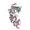
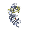
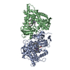
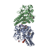
| Title | Crystal Structure of the Plasmid Maintenance System epsilon/zeta: Meachnism of toxin inactivation and toxin function | ||||||
 Components Components |
| ||||||
 Keywords Keywords | POSTSEGREGATIONAL KILLING SYSTEM / PLASMID | ||||||
| Function / homology |  Function and homology information Function and homology informationUDP-N-acetylglucosamine kinase / negative regulation of cell killing / toxic substance binding / response to toxic substance / kinase activity / ATP binding Similarity search - Function | ||||||
| Biological species |  STREPTOCOCCUS PYOGENES (bacteria) STREPTOCOCCUS PYOGENES (bacteria) | ||||||
| Method |  X-RAY DIFFRACTION / SYNCHROTRON / MAD / Resolution: 1.95 Å X-RAY DIFFRACTION / SYNCHROTRON / MAD / Resolution: 1.95 Å | ||||||
 Authors Authors | Meinhart, A. / Alonso, J.C. / Straeter, N. / Saenger, W. | ||||||
 Citation Citation | Journal: Proc.Natl.Acad.Sci.USA / Year: 2003 Title: Crystal Structure of the Plasmid Maintenance System Epsilon /Zeta : Functional Mechanism of Toxin Zeta and Inactivation by Epsilon 2 Zeta 2 Complex Formation Authors: Meinhart, A. / Alonso, J.C. / Strater, N. / Saenger, W. | ||||||
| History |
|
- Structure visualization
| Structure viewer | Molecule: MolmilJmol/JSmol |
|---|
- Downloads & links
-Download
| PDBx/mmCIF format | 1gvn.cif.gz | 164.9 KB | Display | PDBx/mmCIF format |
|---|---|---|---|---|
| PDB format | pdb1gvn.ent.gz | 131.8 KB | Display | PDB format |
| PDBx/mmJSON format | 1gvn.json.gz | Tree view | PDBx/mmJSON format | |
| Others |  Other downloads Other downloads |
-Validation report
| Arichive directory | https://data.pdbj.org/pub/pdb/validation_reports/gv/1gvnftp://data.pdbj.org/pub/pdb/validation_reports/gv/1gvn | HTTPS FTP |
|---|
-Related structure data
| Similar structure data |
|---|


-Links
 PDBj
PDBj- Assembly
| Deposited unit | 
| ||||||||||||
|---|---|---|---|---|---|---|---|---|---|---|---|---|---|
| 1 |
| ||||||||||||
| Unit cell |
| ||||||||||||
| Noncrystallographic symmetry (NCS) | NCS oper:
| ||||||||||||
| Details | THE TETRAMERIC ASSEMBLY CONSISTS OF 2 HETERO DIMERS OFEPSILON AND ZETA (A,C AND B,D) |
-Components
| #1: Protein | Mass: 10734.217 Da / Num. of mol.: 2 / Source method: isolated from a natural source / Details: ENCODED ON PLASMID PSM19035 / Source: (natural) STREPTOCOCCUS PYOGENES (bacteria) / References: UniProt: Q57231#2: Protein | Mass: 32444.969 Da / Num. of mol.: 2 / Source method: isolated from a natural source / Details: ENCODED ON PLASMID PSM19035 / Source: (natural) STREPTOCOCCUS PYOGENES (bacteria) / References: UniProt: Q54944#3: Chemical |   Mass: 96.063 Da / Num. of mol.: 2 / Source method: obtained synthetically / Formula: SO4 Mass: 96.063 Da / Num. of mol.: 2 / Source method: obtained synthetically / Formula: SO4#4: Water | ChemComp-HOH / |  Mass: 18.015 Da / Num. of mol.: 696 / Source method: isolated from a natural source / Formula: H2O Mass: 18.015 Da / Num. of mol.: 696 / Source method: isolated from a natural source / Formula: H2O |
|---|
-Experimental details
-Experiment
| Experiment | Method: X-RAY DIFFRACTION / Number of used crystals: 1 |
|---|
- Sample preparation
| Crystal | Density Matthews: 2.75 Å3/Da / Density % sol: 48 % | ||||||||||||||||||||||||||||||||||||
|---|---|---|---|---|---|---|---|---|---|---|---|---|---|---|---|---|---|---|---|---|---|---|---|---|---|---|---|---|---|---|---|---|---|---|---|---|---|
| Crystal grow | pH: 7 / Details: pH 7.00 | ||||||||||||||||||||||||||||||||||||
| Crystal grow | *PLUS Temperature: 291 K / pH: 7.5 / Method: vapor diffusion, hanging drop / Details: Meinhart, A., (2001) Acta Crystallogr., D57, 745. | ||||||||||||||||||||||||||||||||||||
| Components of the solutions | *PLUS
|
-Data collection
| Diffraction | Mean temperature: 100 K |
|---|---|
| Diffraction source | Source: SYNCHROTRON / Site: EMBL/DESY, HAMBURG  / Beamline: X31 / Wavelength: 1.01629 / Beamline: X31 / Wavelength: 1.01629 |
| Detector | Type: MARRESEARCH / Detector: IMAGE PLATE / Date: Mar 3, 2000 / Details: TOROIDAL MIRROR |
| Radiation | Monochromator: DOUBLE CRYSTAL MONOCHROMATOR / Protocol: SINGLE WAVELENGTH / Monochromatic (M) / Laue (L): M / Scattering type: x-ray |
| Radiation wavelength | Wavelength: 1.01629 Å / Relative weight: 1 |
| Reflection | Resolution: 1.95→19.96 Å / Num. obs: 64215 / % possible obs: 95.5 % / Redundancy: 5 % / Biso Wilson estimate: 8 Å2 / Rmerge(I) obs: 0.051 / Net I/σ(I): 15.5 |
| Reflection shell | Resolution: 1.95→2 Å / Redundancy: 4 % / Rmerge(I) obs: 0.17 / Mean I/σ(I) obs: 5.14 / % possible all: 91.8 |
- Processing
| Software |
| ||||||||||||||||||||||||||||||||||||||||||||||||||||||||||||||||||||||||||||||||
|---|---|---|---|---|---|---|---|---|---|---|---|---|---|---|---|---|---|---|---|---|---|---|---|---|---|---|---|---|---|---|---|---|---|---|---|---|---|---|---|---|---|---|---|---|---|---|---|---|---|---|---|---|---|---|---|---|---|---|---|---|---|---|---|---|---|---|---|---|---|---|---|---|---|---|---|---|---|---|---|---|---|
| Refinement | Method to determine structure: MAD / Resolution: 1.95→19.96 Å / Rfactor Rfree error: 0.004 / Data cutoff high absF: 2 / Isotropic thermal model: RESTRAINED / Cross valid method: THROUGHOUT / σ(F): 0 / Stereochemistry target values: MLF
| ||||||||||||||||||||||||||||||||||||||||||||||||||||||||||||||||||||||||||||||||
| Solvent computation | Solvent model: FLAT MODEL / Bsol: 40.7859 Å2 / ksol: 0.338413 e/Å3 | ||||||||||||||||||||||||||||||||||||||||||||||||||||||||||||||||||||||||||||||||
| Displacement parameters | Biso mean: 21 Å2
| ||||||||||||||||||||||||||||||||||||||||||||||||||||||||||||||||||||||||||||||||
| Refine analyze |
| ||||||||||||||||||||||||||||||||||||||||||||||||||||||||||||||||||||||||||||||||
| Refinement step | Cycle: LAST / Resolution: 1.95→19.96 Å
| ||||||||||||||||||||||||||||||||||||||||||||||||||||||||||||||||||||||||||||||||
| Refine LS restraints |
| ||||||||||||||||||||||||||||||||||||||||||||||||||||||||||||||||||||||||||||||||
| LS refinement shell | Resolution: 1.95→2.07 Å / Rfactor Rfree error: 0.013 / Total num. of bins used: 6
| ||||||||||||||||||||||||||||||||||||||||||||||||||||||||||||||||||||||||||||||||
| Xplor file |
| ||||||||||||||||||||||||||||||||||||||||||||||||||||||||||||||||||||||||||||||||
| Refinement | *PLUS Lowest resolution: 20 Å / Rfactor obs: 0.2 / Rfactor Rfree: 0.235 / Rfactor Rwork: 0.199 | ||||||||||||||||||||||||||||||||||||||||||||||||||||||||||||||||||||||||||||||||
| Solvent computation | *PLUS | ||||||||||||||||||||||||||||||||||||||||||||||||||||||||||||||||||||||||||||||||
| Displacement parameters | *PLUS | ||||||||||||||||||||||||||||||||||||||||||||||||||||||||||||||||||||||||||||||||
| Refine LS restraints | *PLUS
|
