Protocol: SINGLE WAVELENGTH / Monochromatic (M) / Laue (L): M / Scattering type: x-ray
Radiation wavelength
Wavelength: 0.9793 Å / Relative weight: 1
Reflection
Resolution: 2.4→48.3 Å / Num. obs: 60274 / % possible obs: 100 % / Redundancy: 13.1 % / Biso Wilson estimate: 48.26 Å2 / Rmerge(I) obs: 0.155 / Net I/σ(I): 15.4
Reflection shell
Resolution: 2.4→2.46 Å / Redundancy: 11.7 % / Rmerge(I) obs: 1.118 / Mean I/σ(I) obs: 2.7 / % possible all: 100
-
Processing
Software
Name
Version
Classification
XDS
2.10.2
datareduction
XSCALE
datascaling
SHELXDE
phasing
Coot
modelbuilding
BUSTER
2.10.2
refinement
Refinement
Method to determine structure: SAD / Resolution: 2.4→35.14 Å / Cor.coef. Fo:Fc: 0.9288 / Cor.coef. Fo:Fc free: 0.9184 / SU R Cruickshank DPI: 0.23 / Cross valid method: THROUGHOUT / σ(F): 0 / SU R Blow DPI: 0.239 / SU Rfree Blow DPI: 0.184 / SU Rfree Cruickshank DPI: 0.183
Rfactor
Num. reflection
% reflection
Selection details
Rfree
0.2082
881
1.46 %
RANDOM
Rwork
0.1776
-
-
-
obs
0.178
60258
99.99 %
-
Displacement parameters
Biso mean: 56.01 Å2
Baniso -1
Baniso -2
Baniso -3
1-
9.8211 Å2
0 Å2
0 Å2
2-
-
8.2859 Å2
0 Å2
3-
-
-
-18.1069 Å2
Refine analyze
Luzzati coordinate error obs: 0.314 Å
Refinement step
Cycle: 1 / Resolution: 2.4→35.14 Å
Protein
Nucleic acid
Ligand
Solvent
Total
Num. atoms
7528
0
66
509
8103
Refine LS restraints
Refine-ID
Type
Dev ideal
Number
Restraint function
Weight
X-RAY DIFFRACTION
t_bond_d
0.01
7773
HARMONIC
2
X-RAY DIFFRACTION
t_angle_deg
1.03
10549
HARMONIC
2
X-RAY DIFFRACTION
t_dihedral_angle_d
3572
SINUSOIDAL
2
X-RAY DIFFRACTION
t_incorr_chiral_ct
X-RAY DIFFRACTION
t_pseud_angle
X-RAY DIFFRACTION
t_trig_c_planes
224
HARMONIC
2
X-RAY DIFFRACTION
t_gen_planes
1118
HARMONIC
5
X-RAY DIFFRACTION
t_it
7773
HARMONIC
20
X-RAY DIFFRACTION
t_nbd
X-RAY DIFFRACTION
t_omega_torsion
3.24
X-RAY DIFFRACTION
t_other_torsion
2.88
X-RAY DIFFRACTION
t_improper_torsion
X-RAY DIFFRACTION
t_chiral_improper_torsion
991
SEMIHARMONIC
5
X-RAY DIFFRACTION
t_sum_occupancies
X-RAY DIFFRACTION
t_utility_distance
X-RAY DIFFRACTION
t_utility_angle
X-RAY DIFFRACTION
t_utility_torsion
X-RAY DIFFRACTION
t_ideal_dist_contact
9178
SEMIHARMONIC
4
LS refinement shell
Resolution: 2.4→2.46 Å / Total num. of bins used: 20
Rfactor
Num. reflection
% reflection
Rfree
0.2541
66
1.48 %
Rwork
0.2191
4381
-
all
0.2196
4447
-
obs
-
-
99.99 %
Refinement TLS params.
Method: refined / Refine-ID: X-RAY DIFFRACTION
ID
L11 (°2)
L12 (°2)
L13 (°2)
L22 (°2)
L23 (°2)
L33 (°2)
S11 (Å °)
S12 (Å °)
S13 (Å °)
S21 (Å °)
S22 (Å °)
S23 (Å °)
S31 (Å °)
S32 (Å °)
S33 (Å °)
T11 (Å2)
T12 (Å2)
T13 (Å2)
T22 (Å2)
T23 (Å2)
T33 (Å2)
Origin x (Å)
Origin y (Å)
Origin z (Å)
1
0.4459
0.0936
-0.182
0.6386
-0.0869
0.378
-0.0465
0.0233
-0.0902
-0.0747
0.0216
-0.0688
0.0203
-0.0028
0.0249
-0.1922
-0.0057
-0.0111
-0.0821
-0.0114
0.0568
36.731
22.4782
71.5917
2
1.1513
-0.6607
0.3105
3.4109
-0.189
1.2536
-0.1986
-0.3752
-0.1308
1.3019
0.2142
-0.0346
-0.1302
-0.0235
-0.0156
0.4104
0.0833
-0.0401
0.0951
0.0394
-0.0866
29.0638
37.754
115.6
3
0.0515
-0.2633
0.28
0
0.8018
0
0.0001
0.0195
0.0005
-0.0413
0.0015
-0.0193
0.0462
0.0049
-0.0016
-0.0926
0.0967
-0.1749
0.132
-0.06
0.0746
34.5651
19.1555
52.2283
4
0.4131
-0.0656
0.8124
0
-0.2442
0.2124
-0.0096
0.0255
-0.005
0.0038
-0.0045
0.0209
-0.0067
0.0638
0.0142
-0.0517
0.0965
-0.0479
0.1614
-0.0467
-0.0931
28.8283
25.2942
93.3358
5
0
-0.0078
-0.0775
0
0.0484
0
-0.0004
0.0062
-0.0053
0.0153
0.0018
0.0031
0.0088
-0.0006
-0.0014
0.0545
0.0557
0.0431
-0.0578
-0.1091
0.0512
19.2541
29.1125
90.1187
Refinement TLS group
ID
Refine-ID
Refine TLS-ID
Selection details
1
X-RAY DIFFRACTION
1
{A|30 - 501}
2
X-RAY DIFFRACTION
2
{B|30 - 501}
3
X-RAY DIFFRACTION
3
{L|3 - 3}
4
X-RAY DIFFRACTION
4
{L|1 - 1}
5
X-RAY DIFFRACTION
5
{L|2 - 2}
+
About Yorodumi
-
News
-
Feb 9, 2022. New format data for meta-information of EMDB entries
New format data for meta-information of EMDB entries
Version 3 of the EMDB header file is now the official format.
The previous official version 1.9 will be removed from the archive.
In the structure databanks used in Yorodumi, some data are registered as the other names, "COVID-19 virus" and "2019-nCoV". Here are the details of the virus and the list of structure data.
Jan 31, 2019. EMDB accession codes are about to change! (news from PDBe EMDB page)
EMDB accession codes are about to change! (news from PDBe EMDB page)
The allocation of 4 digits for EMDB accession codes will soon come to an end. Whilst these codes will remain in use, new EMDB accession codes will include an additional digit and will expand incrementally as the available range of codes is exhausted. The current 4-digit format prefixed with “EMD-” (i.e. EMD-XXXX) will advance to a 5-digit format (i.e. EMD-XXXXX), and so on. It is currently estimated that the 4-digit codes will be depleted around Spring 2019, at which point the 5-digit format will come into force.
The EM Navigator/Yorodumi systems omit the EMD- prefix.
Related info.:Q: What is EMD? / ID/Accession-code notation in Yorodumi/EM Navigator
Yorodumi is a browser for structure data from EMDB, PDB, SASBDB, etc.
This page is also the successor to EM Navigator detail page, and also detail information page/front-end page for Omokage search.
The word "yorodu" (or yorozu) is an old Japanese word meaning "ten thousand". "mi" (miru) is to see.
Related info.:EMDB / PDB / SASBDB / Comparison of 3 databanks / Yorodumi Search / Aug 31, 2016. New EM Navigator & Yorodumi / Yorodumi Papers / Jmol/JSmol / Function and homology information / Changes in new EM Navigator and Yorodumi
 Movie
Movie Controller

 Yorodumi
 Open data
 Basic information
 Components
 Keywords
 Function and homology information
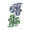
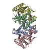
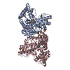
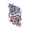
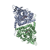
 Porphyromonas gingivalis (bacteria)
 X-RAY DIFFRACTION /
  Authors
 United States,
  Spain, 8items
  Citation
 Structure visualization
 Downloads & links
 Other downloads
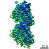


 PDBj
PDBj


 Assembly
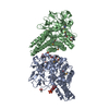

 Type: D-saccharide, beta linking / Mass: 164.156 Da / Num. of mol.: 1
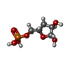
 Type: D-saccharide, beta linking / Mass: 214.110 Da / Num. of mol.: 1 / Source method: obtained synthetically / Formula: C5H11O7P


 Mass: 260.136 Da / Num. of mol.: 1 / Source method: obtained synthetically / Formula: C6H13O9P
 Mass: 92.094 Da / Num. of mol.: 3 / Source method: obtained synthetically / Formula: C3H8O3
 Mass: 59.044 Da / Num. of mol.: 2 / Source method: obtained synthetically / Formula: C2H3O2
 Sample preparation
 Processing