 Movie
Movie Controller


+ Open data

- Basic information
| Entry | Database: PDB / ID: 1g2q | ||||||
|---|---|---|---|---|---|---|---|
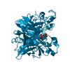
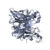
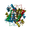
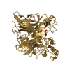
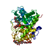
| Title | CRYSTAL STRUCTURE OF ADENINE PHOSPHORIBOSYLTRANSFERASE | ||||||
 Components Components | ADENINE PHOSPHORIBOSYLTRANSFERASE 1 | ||||||
 Keywords Keywords | TRANSFERASE / dimer / single domain / catalytic loop | ||||||
| Function / homology |  Function and homology information Function and homology informationPurine salvage / adenine binding / adenine salvage / adenine phosphoribosyltransferase / adenine phosphoribosyltransferase activity / AMP salvage / purine ribonucleoside salvage / AMP binding / Neutrophil degranulation / metal ion binding ...Purine salvage / adenine binding / adenine salvage / adenine phosphoribosyltransferase / adenine phosphoribosyltransferase activity / AMP salvage / purine ribonucleoside salvage / AMP binding / Neutrophil degranulation / metal ion binding / nucleus / cytoplasm Similarity search - Function | ||||||
| Biological species |  | ||||||
| Method |  X-RAY DIFFRACTION / SYNCHROTRON / MOLECULAR REPLACEMENT / Resolution: 1.5 Å X-RAY DIFFRACTION / SYNCHROTRON / MOLECULAR REPLACEMENT / Resolution: 1.5 Å | ||||||
 Authors Authors | Shi, W. / Tanaka, K.S.E. / Almo, S.C. / Schramm, V.L. | ||||||
 Citation Citation | Journal: Biochemistry / Year: 2001 Title: Structural analysis of adenine phosphoribosyltransferase from Saccharomyces cerevisiae. Authors: Shi, W. / Tanaka, K.S. / Crother, T.R. / Taylor, M.W. / Almo, S.C. / Schramm, V.L. | ||||||
| History |
|
- Structure visualization
| Structure viewer | Molecule: MolmilJmol/JSmol |
|---|
- Downloads & links
-Download
| PDBx/mmCIF format | 1g2q.cif.gz | 86 KB | Display | PDBx/mmCIF format |
|---|---|---|---|---|
| PDB format | pdb1g2q.ent.gz | 64.1 KB | Display | PDB format |
| PDBx/mmJSON format | 1g2q.json.gz | Tree view | PDBx/mmJSON format | |
| Others |  Other downloads Other downloads |
-Validation report
| Arichive directory | https://data.pdbj.org/pub/pdb/validation_reports/g2/1g2qftp://data.pdbj.org/pub/pdb/validation_reports/g2/1g2q | HTTPS FTP |
|---|
-Related structure data
| Related structure data |  1g2pSC S: Starting model for refinement C: citing same article ( |
|---|---|
| Similar structure data |


-Links
 PDBj
PDBj


- Assembly
| Deposited unit | 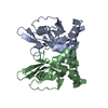
| ||||||||
|---|---|---|---|---|---|---|---|---|---|
| 1 |
| ||||||||
| Unit cell |
| ||||||||
| Details | The biological assembly is a dimer in the asymmetric unit. |
-Components
| #1: Protein | Mass: 20616.814 Da / Num. of mol.: 2 Source method: isolated from a genetically manipulated source Source: (gene. exp.) Plasmid: PQE / Production host:  References: UniProt: P49435, adenine phosphoribosyltransferase #2: Water | ChemComp-HOH / |  Mass: 18.015 Da / Num. of mol.: 305 / Source method: isolated from a natural source / Formula: H2O Mass: 18.015 Da / Num. of mol.: 305 / Source method: isolated from a natural source / Formula: H2O |
|---|
-Experimental details
-Experiment
| Experiment | Method: X-RAY DIFFRACTION / Number of used crystals: 1 |
|---|
- Sample preparation
| Crystal | Density Matthews: 2.18 Å3/Da / Density % sol: 43 % | ||||||||||||||||||||||||
|---|---|---|---|---|---|---|---|---|---|---|---|---|---|---|---|---|---|---|---|---|---|---|---|---|---|
| Crystal grow | Temperature: 298 K / Method: vapor diffusion, hanging drop / pH: 5.5 Details: PEG 5000 MME, sodium acetate, citrate, pH 5.5, VAPOR DIFFUSION, HANGING DROP, temperature 298K | ||||||||||||||||||||||||
| Crystal grow | *PLUS Method: vapor diffusion | ||||||||||||||||||||||||
| Components of the solutions | *PLUS
|
-Data collection
| Diffraction | Mean temperature: 100 K |
|---|---|
| Diffraction source | Source: SYNCHROTRON / Site: NSLS  / Beamline: X9B / Wavelength: 0.98 Å / Beamline: X9B / Wavelength: 0.98 Å |
| Detector | Type: ADSC QUANTUM 4 / Detector: CCD / Date: Feb 14, 2000 |
| Radiation | Protocol: SINGLE WAVELENGTH / Monochromatic (M) / Laue (L): M / Scattering type: x-ray |
| Radiation wavelength | Wavelength: 0.98 Å / Relative weight: 1 |
| Reflection | Resolution: 1.5→20 Å / Num. all: 54848 / Num. obs: 54848 / % possible obs: 95.8 % / Observed criterion σ(F): 0 / Observed criterion σ(I): 0 / Redundancy: 2.4 % / Biso Wilson estimate: 16.1 Å2 / Rsym value: 0.024 / Net I/σ(I): 34.4 |
| Reflection shell | Resolution: 1.5→1.55 Å / Redundancy: 2 % / Mean I/σ(I) obs: 4.3 / Num. unique all: 5137 / Rsym value: 0.176 / % possible all: 89.9 |
| Reflection | *PLUS Num. measured all: 132505 / Rmerge(I) obs: 0.024 |
| Reflection shell | *PLUS % possible obs: 89.9 % / Rmerge(I) obs: 0.176 / Mean I/σ(I) obs: 3.9 |
- Processing
| Software |
| ||||||||||||||||||||||||||||||||||||||||
|---|---|---|---|---|---|---|---|---|---|---|---|---|---|---|---|---|---|---|---|---|---|---|---|---|---|---|---|---|---|---|---|---|---|---|---|---|---|---|---|---|---|
| Refinement | Method to determine structure: MOLECULAR REPLACEMENT Starting model: pdb entry 1G2P Resolution: 1.5→20 Å / Rfactor Rfree error: 0.003 / Isotropic thermal model: restrained / Cross valid method: THROUGHOUT / σ(F): 2 / σ(I): 1.4 / Stereochemistry target values: Engh & Huber
| ||||||||||||||||||||||||||||||||||||||||
| Solvent computation | Solvent model: Flat Model / Bsol: 53.7292 Å2 / ksol: 0.414732 e/Å3 | ||||||||||||||||||||||||||||||||||||||||
| Displacement parameters | Biso mean: 19.3 Å2
| ||||||||||||||||||||||||||||||||||||||||
| Refine analyze |
| ||||||||||||||||||||||||||||||||||||||||
| Refinement step | Cycle: LAST / Resolution: 1.5→20 Å
| ||||||||||||||||||||||||||||||||||||||||
| Refine LS restraints |
| ||||||||||||||||||||||||||||||||||||||||
| LS refinement shell | Resolution: 1.5→1.59 Å / Rfactor Rfree error: 0.009 / Total num. of bins used: 6
| ||||||||||||||||||||||||||||||||||||||||
| Software | *PLUS Name: CNS / Version: 0.9 / Classification: refinement | ||||||||||||||||||||||||||||||||||||||||
| Refinement | *PLUS Lowest resolution: 20 Å / σ(F): 2 / % reflection Rfree: 10.1 % / Rfactor all: 0.202 / Rfactor obs: 0.199 | ||||||||||||||||||||||||||||||||||||||||
| Solvent computation | *PLUS | ||||||||||||||||||||||||||||||||||||||||
| Displacement parameters | *PLUS Biso mean: 19.3 Å2 | ||||||||||||||||||||||||||||||||||||||||
| Refine LS restraints | *PLUS
| ||||||||||||||||||||||||||||||||||||||||
| LS refinement shell | *PLUS Rfactor Rfree: 0.247 / % reflection Rfree: 10.3 % / Rfactor Rwork: 0.231 |
