 Movie
Movie Controller

[English] 日本語
 Yorodumi
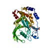
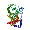
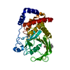
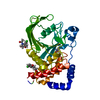
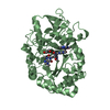
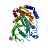
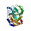
- PDB-1g1h: CRYSTAL STRUCTURE OF PROTEIN TYROSINE PHOSPHATASE 1B COMPLEXED WI... -
+ Open data

- Basic information
| Entry | Database: PDB / ID: 1g1h | ||||||
|---|---|---|---|---|---|---|---|
| Title | CRYSTAL STRUCTURE OF PROTEIN TYROSINE PHOSPHATASE 1B COMPLEXED WITH A BIS-PHOSPHORYLATED PEPTIDE (ETD(PTR)(PTR)RKGGKGLL) FROM THE INSULIN RECEPTOR KINASE | ||||||
 Components Components |
| ||||||
 Keywords Keywords | HYDROLASE / SIGNALING PROTEIN / HYDROLASE (PHOSPHORYLATION) / TYROSINE PHOSPHATASE / PEPTIDE COMPLEX | ||||||
| Function / homology |  Function and homology information Function and homology informationregulation of hepatocyte growth factor receptor signaling pathway / PTK6 Down-Regulation / positive regulation of receptor catabolic process / insulin receptor recycling / peptidyl-tyrosine dephosphorylation involved in inactivation of protein kinase activity / negative regulation of vascular endothelial growth factor receptor signaling pathway / negative regulation of PERK-mediated unfolded protein response / regulation of intracellular protein transport / IRE1-mediated unfolded protein response / cytoplasmic side of endoplasmic reticulum membrane ...regulation of hepatocyte growth factor receptor signaling pathway / PTK6 Down-Regulation / positive regulation of receptor catabolic process / insulin receptor recycling / peptidyl-tyrosine dephosphorylation involved in inactivation of protein kinase activity / negative regulation of vascular endothelial growth factor receptor signaling pathway / negative regulation of PERK-mediated unfolded protein response / regulation of intracellular protein transport / IRE1-mediated unfolded protein response / cytoplasmic side of endoplasmic reticulum membrane / platelet-derived growth factor receptor-beta signaling pathway / sorting endosome / mitochondrial crista / positive regulation of IRE1-mediated unfolded protein response / regulation of type I interferon-mediated signaling pathway / regulation of endocytosis / non-membrane spanning protein tyrosine phosphatase activity / positive regulation of protein tyrosine kinase activity / peptidyl-tyrosine dephosphorylation / Regulation of IFNA/IFNB signaling / regulation of signal transduction / cellular response to unfolded protein / growth hormone receptor signaling pathway via JAK-STAT / negative regulation of endoplasmic reticulum stress-induced intrinsic apoptotic signaling pathway / negative regulation of signal transduction / Regulation of IFNG signaling / MECP2 regulates neuronal receptors and channels / Growth hormone receptor signaling / endoplasmic reticulum unfolded protein response / positive regulation of JUN kinase activity / negative regulation of MAP kinase activity / negative regulation of insulin receptor signaling pathway / Insulin receptor recycling / ephrin receptor binding / Integrin signaling / protein dephosphorylation / protein-tyrosine-phosphatase / protein phosphatase 2A binding / protein tyrosine phosphatase activity / endosome lumen / insulin receptor binding / Negative regulation of MET activity / negative regulation of ERK1 and ERK2 cascade / receptor tyrosine kinase binding / insulin receptor signaling pathway / actin cytoskeleton organization / early endosome / mitochondrial matrix / cadherin binding / protein kinase binding / enzyme binding / endoplasmic reticulum / protein-containing complex / RNA binding / zinc ion binding / cytoplasm / cytosol Similarity search - Function | ||||||
| Biological species |  Homo sapiens (human) Homo sapiens (human) | ||||||
| Method |  X-RAY DIFFRACTION / SYNCHROTRON / Resolution: 2.4 Å X-RAY DIFFRACTION / SYNCHROTRON / Resolution: 2.4 Å | ||||||
 Authors Authors | Salmeen, A. / Andersen, J.N. / Myers, M.P. / Tonks, N.K. / Barford, D. | ||||||
 Citation Citation | Journal: Mol.Cell / Year: 2000 Title: Molecular basis for the dephosphorylation of the activation segment of the insulin receptor by protein tyrosine phosphatase 1B. Authors: Salmeen, A. / Andersen, J.N. / Myers, M.P. / Tonks, N.K. / Barford, D. | ||||||
| History |
|
- Structure visualization
| Structure viewer | Molecule: MolmilJmol/JSmol |
|---|
- Downloads & links
-Download
| PDBx/mmCIF format | 1g1h.cif.gz | 72.9 KB | Display | PDBx/mmCIF format |
|---|---|---|---|---|
| PDB format | pdb1g1h.ent.gz | 57.4 KB | Display | PDB format |
| PDBx/mmJSON format | 1g1h.json.gz | Tree view | PDBx/mmJSON format | |
| Others |  Other downloads Other downloads |
-Validation report
| Summary document | 1g1h_validation.pdf.gz | 428.6 KB | Display | wwPDB validaton report |
|---|---|---|---|---|
| Full document | 1g1h_full_validation.pdf.gz | 429.6 KB | Display | |
| Data in XML | 1g1h_validation.xml.gz | 13.8 KB | Display | |
| Data in CIF | 1g1h_validation.cif.gz | 19.3 KB | Display | |
| Arichive directory | https://data.pdbj.org/pub/pdb/validation_reports/g1/1g1hftp://data.pdbj.org/pub/pdb/validation_reports/g1/1g1h | HTTPS FTP |
-Related structure data


-Links
 PDBj
PDBj


- Assembly
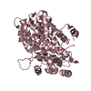
| Deposited unit | 
| ||||||||||
|---|---|---|---|---|---|---|---|---|---|---|---|
| 1 |
| ||||||||||
| Unit cell |
|
-Components
| #1: Protein | Mass: 34688.504 Da / Num. of mol.: 1 / Fragment: CATALYTIC DOMAIN / Mutation: C215A Source method: isolated from a genetically manipulated source Source: (gene. exp.) Homo sapiens (human) / Plasmid: PET-19B / Production host:  |
|---|---|
| #2: Protein/peptide | Mass: 1662.651 Da / Num. of mol.: 1 / Source method: obtained synthetically Details: Sequence from the activation segment of the Insulin Recptor Kinase |
| #3: Water | ChemComp-HOH /  Mass: 18.015 Da / Num. of mol.: 116 / Source method: isolated from a natural source / Formula: H2O Mass: 18.015 Da / Num. of mol.: 116 / Source method: isolated from a natural source / Formula: H2O |
-Experimental details
-Experiment
| Experiment | Method: X-RAY DIFFRACTION / Number of used crystals: 1 |
|---|
- Sample preparation
| Crystal | Density Matthews: 2.94 Å3/Da / Density % sol: 58.14 % | ||||||||||||||||||||||||||||||||||||||||||||||||||||||||
|---|---|---|---|---|---|---|---|---|---|---|---|---|---|---|---|---|---|---|---|---|---|---|---|---|---|---|---|---|---|---|---|---|---|---|---|---|---|---|---|---|---|---|---|---|---|---|---|---|---|---|---|---|---|---|---|---|---|
| Crystal grow | Temperature: 302 K / Method: vapor diffusion, hanging drop / pH: 7.5 Details: PEG 8000, MgCl2, dithiothreitol, HEPES, pH 7.5, VAPOR DIFFUSION, HANGING DROP, temperature 302.0K | ||||||||||||||||||||||||||||||||||||||||||||||||||||||||
| Crystal grow | *PLUS Temperature: 4 ℃ / pH: 7 | ||||||||||||||||||||||||||||||||||||||||||||||||||||||||
| Components of the solutions | *PLUS
|
-Data collection
| Diffraction | Mean temperature: 100 K |
|---|---|
| Diffraction source | Source: SYNCHROTRON / Site: SRS  / Beamline: PX9.6 / Wavelength: 1.488 / Beamline: PX9.6 / Wavelength: 1.488 |
| Detector | Type: MARRESEARCH / Detector: IMAGE PLATE / Date: Mar 17, 1999 |
| Radiation | Protocol: SINGLE WAVELENGTH / Monochromatic (M) / Laue (L): M / Scattering type: x-ray |
| Radiation wavelength | Wavelength: 1.488 Å / Relative weight: 1 |
| Reflection | Resolution: 2.4→30 Å / Num. all: 16411 / Num. obs: 16411 / % possible obs: 99.4 % / Observed criterion σ(F): 0 / Observed criterion σ(I): 0 / Redundancy: 6.4 % / Biso Wilson estimate: 36.4 Å2 / Rmerge(I) obs: 0.058 / Net I/σ(I): 20.3 |
| Reflection shell | Highest resolution: 2.4 Å / Redundancy: 3.1 % / Rmerge(I) obs: 0.132 / Num. unique all: 1601 / % possible all: 96 |
| Reflection | *PLUS Num. measured all: 105145 |
| Reflection shell | *PLUS % possible obs: 96 % |
- Processing
| Software |
| |||||||||||||||||||||||||
|---|---|---|---|---|---|---|---|---|---|---|---|---|---|---|---|---|---|---|---|---|---|---|---|---|---|---|
| Refinement | Resolution: 2.4→30 Å / Cross valid method: THROUGHOUT / σ(F): 0 / σ(I): 0 / Stereochemistry target values: CNS
| |||||||||||||||||||||||||
| Refinement step | Cycle: LAST / Resolution: 2.4→30 Å
| |||||||||||||||||||||||||
| Refine LS restraints |
| |||||||||||||||||||||||||
| Software | *PLUS Name: CNS / Classification: refinement | |||||||||||||||||||||||||
| Refinement | *PLUS Lowest resolution: 30 Å / σ(F): 0 / % reflection Rfree: 5 % / Rfactor obs: 0.207 | |||||||||||||||||||||||||
| Solvent computation | *PLUS | |||||||||||||||||||||||||
| Displacement parameters | *PLUS | |||||||||||||||||||||||||
| Refine LS restraints | *PLUS Type: c_angle_deg / Dev ideal: 2.5 |
