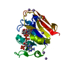
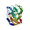
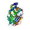
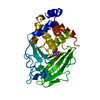
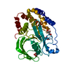
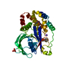
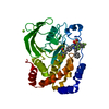
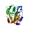
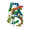
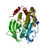
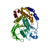
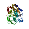
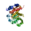
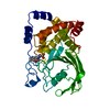
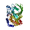
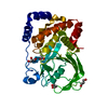
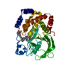
Entry Database : PDB / ID : 7mm1Title PTP1B in complex with TCS401 by Native S-SAD at Room Temperature Tyrosine-protein phosphatase non-receptor type 1 Keywords / / / / Function / homology Function Domain/homology Component
/ / / / / / / / / / / / / / / / / / / / / / / / / / / / / / / / / / / / / / / / / / / / / / / / / / / / / / / / / / / / / / / / / / / / / / / / / / / / / / / / / / / / / / / / / / / / / / / / / / / / / / / / / / Biological species Homo sapiens (human)Method / / / Resolution : 1.85 Å Authors Greisman, J.B. / Dalton, K.M. / Hekstra, D.R. Funding support Organization Grant number Country Other private Searle Scholars Program (SSP-2018-3240) Other private George W. Merck Fund in the New York Community Trust (338034) National Science Foundation (NSF, United States) DGE1745303
Journal : Acta Crystallogr D Struct Biol / Year : 2022Title : Native SAD phasing at room temperature.Authors : Greisman, J.B. / Dalton, K.M. / Sheehan, C.J. / Klureza, M.A. / Kurinov, I. / Hekstra, D.R. History Deposition Apr 29, 2021 Deposition site / Processing site Revision 1.0 May 12, 2021 Provider / Type Revision 1.1 May 26, 2021 Group / Category Revision 1.2 Aug 3, 2022 Group / Category / citation_author / database_2Item _citation.country / _citation.journal_abbrev ... _citation.country / _citation.journal_abbrev / _citation.journal_id_ASTM / _citation.journal_id_CSD / _citation.journal_id_ISSN / _citation.journal_volume / _citation.pdbx_database_id_DOI / _citation.title / _citation.year / _database_2.pdbx_DOI / _database_2.pdbx_database_accession Revision 1.3 Aug 17, 2022 Group / Category / citation_authorItem _citation.country / _citation.journal_abbrev ... _citation.country / _citation.journal_abbrev / _citation.journal_id_ASTM / _citation.journal_id_ISSN / _citation.page_first / _citation.page_last / _citation.pdbx_database_id_PubMed / _citation.title / _citation_author.identifier_ORCID Revision 1.4 May 22, 2024 Group / Category / chem_comp_bond
Show all Show less
 Movie
Movie Controller


 Open data
 Basic information
 Components
 Keywords
 Function and homology information
 Homo sapiens (human)
 X-RAY DIFFRACTION /
  Authors
 United States, 3items
  Citation
 Structure visualization
 Downloads & links
 Other downloads
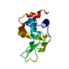


 PDBj
PDBj


 Assembly
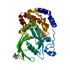

 Mass: 270.262 Da / Num. of mol.: 1 / Source method: obtained synthetically / Formula: C10H10N2O5S
 Mass: 122.143 Da / Num. of mol.: 2 / Source method: obtained synthetically / Formula: C4H12NO3 / Comment: pH buffer*YM
 Mass: 18.015 Da / Num. of mol.: 159 / Source method: isolated from a natural source / Formula: H2O
 Sample preparation
 Processing