 Movie
Movie Controller

[English] 日本語
 Yorodumi
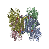
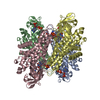
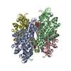
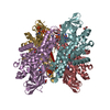
- PDB-1egc: STRUCTURE OF T255E, E376G MUTANT OF HUMAN MEDIUM CHAIN ACYL-COA D... -
+ Open data

- Basic information
| Entry | Database: PDB / ID: 1egc | ||||||
|---|---|---|---|---|---|---|---|
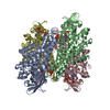
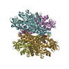
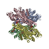
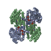
| Title | STRUCTURE OF T255E, E376G MUTANT OF HUMAN MEDIUM CHAIN ACYL-COA DEHYDROGENASE COMPLEXED WITH OCTANOYL-COA | ||||||
 Components Components | MEDIUM CHAIN ACYL-COA DEHYDROGENASE | ||||||
 Keywords Keywords | ELECTRON TRANSFER / ACYL-COA DEHYDROGENASE / FLAVOPROTEIN | ||||||
| Function / homology |  Function and homology information Function and homology informationmedium-chain fatty acid catabolic process / mitochondrial fatty acid beta-oxidation of unsaturated fatty acids / carnitine metabolic process, CoA-linked / medium-chain acyl-CoA dehydrogenase / medium-chain fatty acyl-CoA dehydrogenase activity / Beta oxidation of octanoyl-CoA to hexanoyl-CoA / Beta oxidation of decanoyl-CoA to octanoyl-CoA-CoA / carnitine biosynthetic process / medium-chain fatty acid metabolic process / fatty acid beta-oxidation using acyl-CoA dehydrogenase ...medium-chain fatty acid catabolic process / mitochondrial fatty acid beta-oxidation of unsaturated fatty acids / carnitine metabolic process, CoA-linked / medium-chain acyl-CoA dehydrogenase / medium-chain fatty acyl-CoA dehydrogenase activity / Beta oxidation of octanoyl-CoA to hexanoyl-CoA / Beta oxidation of decanoyl-CoA to octanoyl-CoA-CoA / carnitine biosynthetic process / medium-chain fatty acid metabolic process / fatty acid beta-oxidation using acyl-CoA dehydrogenase / acyl-CoA dehydrogenase activity / cardiac muscle cell differentiation / glycogen biosynthetic process / regulation of gluconeogenesis / response to starvation / fatty acid beta-oxidation / response to cold / post-embryonic development / liver development / PPARA activates gene expression / mitochondrial membrane / flavin adenine dinucleotide binding / mitochondrial matrix / axon / mitochondrion / identical protein binding / nucleus / cytoplasm Similarity search - Function | ||||||
| Biological species |  Homo sapiens (human) Homo sapiens (human) | ||||||
| Method |  X-RAY DIFFRACTION / Resolution: 2.6 Å X-RAY DIFFRACTION / Resolution: 2.6 Å | ||||||
 Authors Authors | Lee, H.J. / Wang, M. / Paschke, R. / Nandy, A. / Ghisla, S. / Kim, J.P. | ||||||
 Citation Citation | Journal: Biochemistry / Year: 1996 Title: Crystal structures of the wild type and the Glu376Gly/Thr255Glu mutant of human medium-chain acyl-CoA dehydrogenase: influence of the location of the catalytic base on substrate specificity. Authors: Lee, H.J. / Wang, M. / Paschke, R. / Nandy, A. / Ghisla, S. / Kim, J.J. | ||||||
| History |
|
- Structure visualization
| Structure viewer | Molecule: MolmilJmol/JSmol |
|---|
- Downloads & links
-Download
| PDBx/mmCIF format | 1egc.cif.gz | 310.1 KB | Display | PDBx/mmCIF format |
|---|---|---|---|---|
| PDB format | pdb1egc.ent.gz | 254.1 KB | Display | PDB format |
| PDBx/mmJSON format | 1egc.json.gz | Tree view | PDBx/mmJSON format | |
| Others |  Other downloads Other downloads |
-Validation report
| Arichive directory | https://data.pdbj.org/pub/pdb/validation_reports/eg/1egcftp://data.pdbj.org/pub/pdb/validation_reports/eg/1egc | HTTPS FTP |
|---|
-Related structure data


-Links
 PDBj
PDBj- Assembly
| Deposited unit | 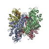
| ||||||||
|---|---|---|---|---|---|---|---|---|---|
| 1 |
| ||||||||
| Unit cell |
|
-Components
| #1: Protein | Mass: 43651.762 Da / Num. of mol.: 4 / Mutation: T255E, E376G Source method: isolated from a genetically manipulated source Details: COMPLEXED WITH OCTANOYL-COA / Source: (gene. exp.) Homo sapiens (human) / References: UniProt: P11310, EC: 1.3.99.3#2: Chemical | ChemComp-CO8 / 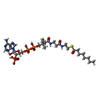  Mass: 893.730 Da / Num. of mol.: 4 / Source method: obtained synthetically / Formula: C29H50N7O17P3S Mass: 893.730 Da / Num. of mol.: 4 / Source method: obtained synthetically / Formula: C29H50N7O17P3S#3: Chemical | ChemComp-FAD /   Mass: 785.550 Da / Num. of mol.: 4 / Source method: obtained synthetically / Formula: C27H33N9O15P2 / Comment: FAD*YM Mass: 785.550 Da / Num. of mol.: 4 / Source method: obtained synthetically / Formula: C27H33N9O15P2 / Comment: FAD*YM#4: Water | ChemComp-HOH / |  Mass: 18.015 Da / Num. of mol.: 136 / Source method: isolated from a natural source / Formula: H2O Mass: 18.015 Da / Num. of mol.: 136 / Source method: isolated from a natural source / Formula: H2O |
|---|
-Experimental details
-Experiment
| Experiment | Method: X-RAY DIFFRACTION |
|---|
- Sample preparation
| Crystal | Density Matthews: 3.1 Å3/Da / Density % sol: 49 % | ||||||||||||||||||||||||||||||
|---|---|---|---|---|---|---|---|---|---|---|---|---|---|---|---|---|---|---|---|---|---|---|---|---|---|---|---|---|---|---|---|
| Crystal grow | *PLUS Temperature: 4 ℃ / pH: 7 / Method: vapor diffusion, sitting drop | ||||||||||||||||||||||||||||||
| Components of the solutions | *PLUS
|
-Data collection
| Diffraction source | Wavelength: 1.5418 |
|---|---|
| Detector | Type: RIGAKU RAXIS IIC / Detector: IMAGE PLATE / Date: Mar 8, 1994 |
| Radiation | Monochromatic (M) / Laue (L): M / Scattering type: x-ray |
| Radiation wavelength | Wavelength: 1.5418 Å / Relative weight: 1 |
| Reflection | Num. obs: 51243 / % possible obs: 70.7 % / Observed criterion σ(I): 1 / Redundancy: 2.7 % / Rmerge(I) obs: 0.101 |
| Reflection | *PLUS Highest resolution: 2.6 Å / Num. measured all: 137635 / Rmerge(I) obs: 0.1018 |
- Processing
| Software |
| ||||||||||||||||||||||||||||||||||||||||||||||||||||||||||||
|---|---|---|---|---|---|---|---|---|---|---|---|---|---|---|---|---|---|---|---|---|---|---|---|---|---|---|---|---|---|---|---|---|---|---|---|---|---|---|---|---|---|---|---|---|---|---|---|---|---|---|---|---|---|---|---|---|---|---|---|---|---|
| Refinement | Resolution: 2.6→10 Å / σ(F): 2 /
| ||||||||||||||||||||||||||||||||||||||||||||||||||||||||||||
| Displacement parameters | Biso mean: 16.16 Å2 | ||||||||||||||||||||||||||||||||||||||||||||||||||||||||||||
| Refinement step | Cycle: LAST / Resolution: 2.6→10 Å
| ||||||||||||||||||||||||||||||||||||||||||||||||||||||||||||
| Refine LS restraints |
| ||||||||||||||||||||||||||||||||||||||||||||||||||||||||||||
| Software | *PLUS Name: X-PLOR / Classification: refinement | ||||||||||||||||||||||||||||||||||||||||||||||||||||||||||||
| Refinement | *PLUS | ||||||||||||||||||||||||||||||||||||||||||||||||||||||||||||
| Solvent computation | *PLUS | ||||||||||||||||||||||||||||||||||||||||||||||||||||||||||||
| Displacement parameters | *PLUS Biso mean: 16.162 Å2 | ||||||||||||||||||||||||||||||||||||||||||||||||||||||||||||
| Refine LS restraints | *PLUS
|
