Mass: 18.015 Da / Num. of mol.: 777 / Source method: isolated from a natural source / Formula: H2O
-
Experimental details
-
Experiment
Experiment
Method: X-RAY DIFFRACTION / Number of used crystals: 1
-
Sample preparation
Crystal
Density Matthews: 2.29 Å3/Da / Density % sol: 46.32 %
Crystal grow
Temperature: 277 K / Method: vapor diffusion, hanging drop / pH: 8.5 Details: 1 mM FAD, 50% (v,v) MPD, 0.1 M Tris/HCl, pH 8.5 and 0.2 M NH4H2PO4, VAPOR DIFFUSION, HANGING DROP, temperature 277K
Resolution: 2.1→30 Å / Cor.coef. Fo:Fc: 0.955 / Cor.coef. Fo:Fc free: 0.931 / SU B: 12.712 / SU ML: 0.147 / Cross valid method: THROUGHOUT / ESU R: 0.224 / ESU R Free: 0.182 / Stereochemistry target values: MAXIMUM LIKELIHOOD / Details: HYDROGENS HAVE BEEN USED IF PRESENT IN THE INPUT
Rfactor
Num. reflection
% reflection
Selection details
Rfree
0.22466
7062
5 %
RANDOM
Rwork
0.18094
-
-
-
obs
0.18317
133695
99.75 %
-
all
-
133695
-
-
Solvent computation
Ion probe radii: 0.8 Å / Shrinkage radii: 0.8 Å / VDW probe radii: 1.2 Å / Solvent model: MASK
Displacement parameters
Biso mean: 38.967 Å2
Baniso -1
Baniso -2
Baniso -3
1-
0.59 Å2
0 Å2
0 Å2
2-
-
0.35 Å2
0 Å2
3-
-
-
-0.95 Å2
Refinement step
Cycle: LAST / Resolution: 2.1→30 Å
Protein
Nucleic acid
Ligand
Solvent
Total
Num. atoms
17986
0
324
777
19087
Refine LS restraints
Refine-ID
Type
Dev ideal
Dev ideal target
Number
X-RAY DIFFRACTION
r_bond_refined_d
0.017
0.022
18785
X-RAY DIFFRACTION
r_bond_other_d
X-RAY DIFFRACTION
r_angle_refined_deg
1.641
1.981
25426
X-RAY DIFFRACTION
r_angle_other_deg
X-RAY DIFFRACTION
r_dihedral_angle_1_deg
5.804
5
2361
X-RAY DIFFRACTION
r_dihedral_angle_2_deg
41.025
24.418
833
X-RAY DIFFRACTION
r_dihedral_angle_3_deg
16.867
15
3131
X-RAY DIFFRACTION
r_dihedral_angle_4_deg
20.326
15
105
X-RAY DIFFRACTION
r_chiral_restr
0.125
0.2
2699
X-RAY DIFFRACTION
r_gen_planes_refined
0.008
0.021
14305
X-RAY DIFFRACTION
r_gen_planes_other
X-RAY DIFFRACTION
r_nbd_refined
X-RAY DIFFRACTION
r_nbd_other
X-RAY DIFFRACTION
r_nbtor_refined
X-RAY DIFFRACTION
r_nbtor_other
X-RAY DIFFRACTION
r_xyhbond_nbd_refined
X-RAY DIFFRACTION
r_xyhbond_nbd_other
X-RAY DIFFRACTION
r_metal_ion_refined
X-RAY DIFFRACTION
r_metal_ion_other
X-RAY DIFFRACTION
r_symmetry_vdw_refined
X-RAY DIFFRACTION
r_symmetry_vdw_other
X-RAY DIFFRACTION
r_symmetry_hbond_refined
X-RAY DIFFRACTION
r_symmetry_hbond_other
X-RAY DIFFRACTION
r_symmetry_metal_ion_refined
X-RAY DIFFRACTION
r_symmetry_metal_ion_other
X-RAY DIFFRACTION
r_mcbond_it
0.886
1.5
11628
X-RAY DIFFRACTION
r_mcbond_other
X-RAY DIFFRACTION
r_mcangle_it
1.468
2
18451
X-RAY DIFFRACTION
r_scbond_it
2.932
3
7086
X-RAY DIFFRACTION
r_scangle_it
4.294
4.5
6889
X-RAY DIFFRACTION
r_rigid_bond_restr
X-RAY DIFFRACTION
r_sphericity_free
X-RAY DIFFRACTION
r_sphericity_bonded
Refine LS restraints NCS
Ens-ID: 1 / Refine-ID: X-RAY DIFFRACTION
Dom-ID
Auth asym-ID
Number
Type
Rms dev position (Å)
Weight position
1
B
1536
MEDIUMPOSITIONAL
0.12
0.5
2
A
1536
MEDIUMPOSITIONAL
0.12
0.5
3
D
1536
MEDIUMPOSITIONAL
0.15
0.5
4
E
1536
MEDIUMPOSITIONAL
0.16
0.5
5
F
1536
MEDIUMPOSITIONAL
0.12
0.5
6
G
1536
MEDIUMPOSITIONAL
0.13
0.5
1
B
1445
LOOSEPOSITIONAL
0.3
5
2
A
1445
LOOSEPOSITIONAL
0.27
5
3
D
1445
LOOSEPOSITIONAL
0.33
5
4
E
1445
LOOSEPOSITIONAL
0.39
5
5
F
1445
LOOSEPOSITIONAL
0.3
5
6
G
1445
LOOSEPOSITIONAL
0.37
5
1
B
1536
MEDIUMTHERMAL
1.88
2
2
A
1536
MEDIUMTHERMAL
1.84
2
3
D
1536
MEDIUMTHERMAL
1.93
2
4
E
1536
MEDIUMTHERMAL
2.17
2
5
F
1536
MEDIUMTHERMAL
1.69
2
6
G
1536
MEDIUMTHERMAL
1.97
2
1
B
1445
LOOSETHERMAL
2.59
10
2
A
1445
LOOSETHERMAL
2.78
10
3
D
1445
LOOSETHERMAL
2.73
10
4
E
1445
LOOSETHERMAL
3.17
10
5
F
1445
LOOSETHERMAL
2.52
10
6
G
1445
LOOSETHERMAL
2.76
10
LS refinement shell
Resolution: 2.1→2.154 Å / Total num. of bins used: 20
Rfactor
Num. reflection
% reflection
Rfree
0.287
462
-
Rwork
0.249
9552
-
obs
-
-
97.95 %
Refinement TLS params.
Method: refined / Refine-ID: X-RAY DIFFRACTION
ID
L11 (°2)
L12 (°2)
L13 (°2)
L22 (°2)
L23 (°2)
L33 (°2)
S11 (Å °)
S12 (Å °)
S13 (Å °)
S21 (Å °)
S22 (Å °)
S23 (Å °)
S31 (Å °)
S32 (Å °)
S33 (Å °)
T11 (Å2)
T12 (Å2)
T13 (Å2)
T22 (Å2)
T23 (Å2)
T33 (Å2)
Origin x (Å)
Origin y (Å)
Origin z (Å)
1
0.9189
0.2225
-0.2517
0.8238
-0.3553
1.3272
-0.0207
-0.1331
-0.1677
0.0287
0.0226
0.125
0.148
-0.2034
-0.0019
0.0452
-0.0275
0.0255
0.1459
0.0222
0.0744
56.899
108.461
40.958
2
0.9772
-0.2277
-0.3255
0.9589
0.2691
1.5509
-0.0194
0.0319
-0.2243
-0.092
-0.0002
-0.1842
0.2133
0.2048
0.0196
0.0724
0.055
0.0434
0.0959
0.006
0.122
94.434
107.309
23.269
3
0.9154
-0.191
0.217
1.7696
-0.9476
1.6237
-0.263
0.1348
0.3933
0.4868
-0.1601
-0.3715
-0.6563
0.3288
0.4232
0.4643
-0.1594
-0.3437
0.132
0.17
0.347
75.784
68.559
3.174
4
0.8144
-0.2017
0.1617
1.7536
-1.1432
2.4655
-0.1945
-0.2177
0.0447
0.7184
0.4285
0.5431
-0.7708
-0.6827
-0.2341
0.4605
0.2895
0.1317
0.3052
0.0936
0.2747
44.6
52.244
20.592
5
0.6146
-0.1672
0.4448
1.1808
-0.0265
1.8371
0.0906
0.001
-0.1887
-0.0759
-0.0006
0.21
0.333
-0.0026
-0.09
0.1588
0.0154
-0.0519
0.0709
0.0467
0.1658
62.456
19.168
3.269
6
1.2963
-0.3702
1.0101
0.637
-0.4661
1.8357
-0.1521
0.1187
0.1715
0.2149
-0.1455
-0.3195
-0.1963
0.4684
0.2977
0.1785
-0.0425
-0.1594
0.2913
0.1748
0.2409
93.098
35.728
21.283
Refinement TLS group
ID
Refine-ID
Refine TLS-ID
Auth asym-ID
Auth seq-ID
1
X-RAY DIFFRACTION
1
A
1 - 389
2
X-RAY DIFFRACTION
1
A
400 - 401
3
X-RAY DIFFRACTION
2
B
1 - 393
4
X-RAY DIFFRACTION
2
B
400 - 401
5
X-RAY DIFFRACTION
3
D
1 - 390
6
X-RAY DIFFRACTION
3
D
400 - 401
7
X-RAY DIFFRACTION
4
E
1 - 391
8
X-RAY DIFFRACTION
4
E
400 - 401
9
X-RAY DIFFRACTION
5
F
1 - 390
10
X-RAY DIFFRACTION
5
F
400 - 401
11
X-RAY DIFFRACTION
6
G
1 - 389
12
X-RAY DIFFRACTION
6
G
400 - 401
+
About Yorodumi
-
News
-
Feb 9, 2022. New format data for meta-information of EMDB entries
New format data for meta-information of EMDB entries
Version 3 of the EMDB header file is now the official format.
The previous official version 1.9 will be removed from the archive.
In the structure databanks used in Yorodumi, some data are registered as the other names, "COVID-19 virus" and "2019-nCoV". Here are the details of the virus and the list of structure data.
Jan 31, 2019. EMDB accession codes are about to change! (news from PDBe EMDB page)
EMDB accession codes are about to change! (news from PDBe EMDB page)
The allocation of 4 digits for EMDB accession codes will soon come to an end. Whilst these codes will remain in use, new EMDB accession codes will include an additional digit and will expand incrementally as the available range of codes is exhausted. The current 4-digit format prefixed with “EMD-” (i.e. EMD-XXXX) will advance to a 5-digit format (i.e. EMD-XXXXX), and so on. It is currently estimated that the 4-digit codes will be depleted around Spring 2019, at which point the 5-digit format will come into force.
The EM Navigator/Yorodumi systems omit the EMD- prefix.
Related info.:Q: What is EMD? / ID/Accession-code notation in Yorodumi/EM Navigator
Yorodumi is a browser for structure data from EMDB, PDB, SASBDB, etc.
This page is also the successor to EM Navigator detail page, and also detail information page/front-end page for Omokage search.
The word "yorodu" (or yorozu) is an old Japanese word meaning "ten thousand". "mi" (miru) is to see.
Related info.:EMDB / PDB / SASBDB / Comparison of 3 databanks / Yorodumi Search / Aug 31, 2016. New EM Navigator & Yorodumi / Yorodumi Papers / Jmol/JSmol / Function and homology information / Changes in new EM Navigator and Yorodumi
 Movie
Movie Controller


 Open data
 Basic information
 Components
 Keywords
 Function and homology information
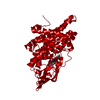
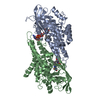
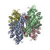
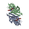
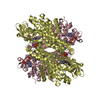
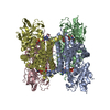
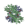
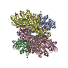
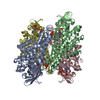
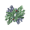
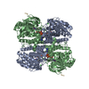
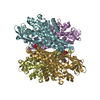
 Desulfococcus multivorans (bacteria)
 X-RAY DIFFRACTION /
  Authors
 Citation
 Structure visualization
 Downloads & links
 Other downloads
 PDBj
PDBj Assembly
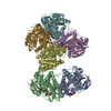

 Mass: 785.550 Da / Num. of mol.: 6 / Source method: obtained synthetically / Formula: C27H33N9O15P2 / Comment: FAD*YM
 Mass: 35.453 Da / Num. of mol.: 6 / Source method: obtained synthetically / Formula: Cl
 Mass: 18.015 Da / Num. of mol.: 777 / Source method: isolated from a natural source / Formula: H2O
 Sample preparation
 / Beamline: X10SA / Wavelength: 1 Å
 Processing