 Movie
Movie Controller

[English] 日本語
 Yorodumi
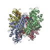
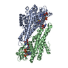
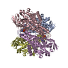
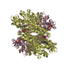
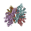
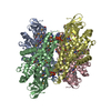
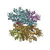
- PDB-1ivh: STRUCTURE OF HUMAN ISOVALERYL-COA DEHYDROGENASE AT 2.6 ANGSTROMS ... -
+ Open data

- Basic information
| Entry | Database: PDB / ID: 1ivh | ||||||
|---|---|---|---|---|---|---|---|
| Title | STRUCTURE OF HUMAN ISOVALERYL-COA DEHYDROGENASE AT 2.6 ANGSTROMS RESOLUTION: STRUCTURAL BASIS FOR SUBSTRATE SPECIFICITY | ||||||
 Components Components | ISOVALERYL-COA DEHYDROGENASE | ||||||
 Keywords Keywords | OXIDOREDUCTASE / ACYL-COA DEHYDROGENASE / FLAVOPROTEIN / ISOVALERYL-COA / ISOVALERIC ACIDEMIA | ||||||
| Function / homology |  Function and homology information Function and homology informationIsovaleric acidemia / isovaleryl-CoA dehydrogenase / 3-methylbutanoyl-CoA dehydrogenase activity / short-chain acyl-CoA dehydrogenase / fatty acid beta-oxidation using acyl-CoA dehydrogenase / L-leucine catabolic process / Branched-chain amino acid catabolism / branched-chain amino acid catabolic process / flavin adenine dinucleotide binding / mitochondrial matrix ...Isovaleric acidemia / isovaleryl-CoA dehydrogenase / 3-methylbutanoyl-CoA dehydrogenase activity / short-chain acyl-CoA dehydrogenase / fatty acid beta-oxidation using acyl-CoA dehydrogenase / L-leucine catabolic process / Branched-chain amino acid catabolism / branched-chain amino acid catabolic process / flavin adenine dinucleotide binding / mitochondrial matrix / mitochondrion / nucleoplasm / identical protein binding Similarity search - Function | ||||||
| Biological species |  Homo sapiens (human) Homo sapiens (human) | ||||||
| Method |  X-RAY DIFFRACTION / MOLECULAR REPLACEMENT / Resolution: 2.6 Å X-RAY DIFFRACTION / MOLECULAR REPLACEMENT / Resolution: 2.6 Å | ||||||
 Authors Authors | Tiffany, K.A. / Roberts, D.L. / Wang, M. / Paschke, R. / Mohsen, A.-W.A. / Vockley, J. / Kim, J.J.P. | ||||||
 Citation Citation | Journal: Biochemistry / Year: 1997 Title: Structure of human isovaleryl-CoA dehydrogenase at 2.6 A resolution: structural basis for substrate specificity,. Authors: Tiffany, K.A. / Roberts, D.L. / Wang, M. / Paschke, R. / Mohsen, A.W. / Vockley, J. / Kim, J.J. | ||||||
| History |
|
- Structure visualization
| Structure viewer | Molecule: MolmilJmol/JSmol |
|---|
- Downloads & links
-Download
| PDBx/mmCIF format | 1ivh.cif.gz | 308.5 KB | Display | PDBx/mmCIF format |
|---|---|---|---|---|
| PDB format | pdb1ivh.ent.gz | 252.7 KB | Display | PDB format |
| PDBx/mmJSON format | 1ivh.json.gz | Tree view | PDBx/mmJSON format | |
| Others |  Other downloads Other downloads |
-Validation report
| Arichive directory | https://data.pdbj.org/pub/pdb/validation_reports/iv/1ivhftp://data.pdbj.org/pub/pdb/validation_reports/iv/1ivh | HTTPS FTP |
|---|
-Related structure data
| Related structure data |  1mdeS S: Starting model for refinement |
|---|---|
| Similar structure data |

-Links
 PDBj
PDBj- Assembly
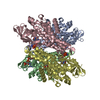
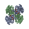
| Deposited unit | 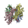
| ||||||||
|---|---|---|---|---|---|---|---|---|---|
| 1 |
| ||||||||
| Unit cell |
|
-Components
| #1: Protein | Mass: 43120.477 Da / Num. of mol.: 4 Source method: isolated from a genetically manipulated source Details: EACH SUBUNIT CONTAINS ONE NON-COVALENTLY BOUND FAD MOLECULE AND ONE NON-COVALENTLY BOUND COA PER SULFIDE MOLECULE Source: (gene. exp.) Homo sapiens (human)Description: THE CDNA WAS ALTERED TO ACCOMMODATE ESCHERICHIA COLI CODON USAGE IN ORDER TO ACHIEVE A HIGH LEVEL OF EXPRESSION. THE MOLECULE WAS THEN CLONED, EXPRESSED, AND PURIFIED AS DESCRIBED IN\: ...Description: THE CDNA WAS ALTERED TO ACCOMMODATE ESCHERICHIA COLI CODON USAGE IN ORDER TO ACHIEVE A HIGH LEVEL OF EXPRESSION. THE MOLECULE WAS THEN CLONED, EXPRESSED, AND PURIFIED AS DESCRIBED IN\: MOHSEN, A.W. AND VOCKLEY, J. (1995) BIOCHEMISTRY, 34\:10146-10152 Gene: IVD / Organ: LIVER / Organelle: MITOCHONDRIA / Plasmid: PKMHIVD / Cellular location (production host): CYTOPLASM / Production host:  #2: Chemical | ChemComp-FAD /   Mass: 785.550 Da / Num. of mol.: 4 / Source method: obtained synthetically / Formula: C27H33N9O15P2 / Comment: FAD*YM Mass: 785.550 Da / Num. of mol.: 4 / Source method: obtained synthetically / Formula: C27H33N9O15P2 / Comment: FAD*YM#3: Chemical | ChemComp-COS / 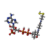  Mass: 799.599 Da / Num. of mol.: 4 / Source method: obtained synthetically / Formula: C21H36N7O16P3S2 Mass: 799.599 Da / Num. of mol.: 4 / Source method: obtained synthetically / Formula: C21H36N7O16P3S2#4: Water | ChemComp-HOH / |  Mass: 18.015 Da / Num. of mol.: 215 / Source method: isolated from a natural source / Formula: H2O Mass: 18.015 Da / Num. of mol.: 215 / Source method: isolated from a natural source / Formula: H2OCompound details | COA PERSULFIDE | Has protein modification | Y | |
|---|
-Experimental details
-Experiment
| Experiment | Method: X-RAY DIFFRACTION / Number of used crystals: 2 |
|---|
- Sample preparation
| Crystal | Density Matthews: 2.4 Å3/Da / Density % sol: 51 % / Description: A HOMOTETRAMER WAS USED AS THE SEARCH MODEL | ||||||||||||||||||||||||||||||
|---|---|---|---|---|---|---|---|---|---|---|---|---|---|---|---|---|---|---|---|---|---|---|---|---|---|---|---|---|---|---|---|
| Crystal grow | Temperature: 292 K / pH: 8.5 Details: PROTEIN WAS CRYSTALLIZED FROM 8% PEG 8000, 0.1 M TRIS, PH 8.5 AT 19 DEGREES CELSIUS., temperature 292K | ||||||||||||||||||||||||||||||
| Crystal grow | *PLUS Temperature: 19 ℃ / Method: vapor diffusion, hanging drop | ||||||||||||||||||||||||||||||
| Components of the solutions | *PLUS
|
-Data collection
| Diffraction | Mean temperature: 277 K |
|---|---|
| Diffraction source | Source: ROTATING ANODE / Type: RIGAKU RUH2R / Wavelength: 1.5418 |
| Detector | Type: RIGAKU / Detector: IMAGE PLATE / Date: Apr 1, 1994 / Details: COLLIMATOR |
| Radiation | Monochromator: GRAPHITE(002) / Monochromatic (M) / Laue (L): M / Scattering type: x-ray |
| Radiation wavelength | Wavelength: 1.5418 Å / Relative weight: 1 |
| Reflection | Resolution: 2.6→30 Å / Num. obs: 42529 / % possible obs: 82.2 % / Observed criterion σ(I): 0 / Redundancy: 2.4 % / Rmerge(I) obs: 0.07 / Rsym value: 0.07 / Net I/σ(I): 11.5 |
| Reflection shell | Resolution: 2.6→2.64 Å / Redundancy: 2.1 % / Mean I/σ(I) obs: 2.3 / Rsym value: 0.219 / % possible all: 61 |
| Reflection shell | *PLUS % possible obs: 61 % / Rmerge(I) obs: 0.219 |
- Processing
| Software |
| ||||||||||||||||||||||||||||||||||||||||||||||||||||||||||||
|---|---|---|---|---|---|---|---|---|---|---|---|---|---|---|---|---|---|---|---|---|---|---|---|---|---|---|---|---|---|---|---|---|---|---|---|---|---|---|---|---|---|---|---|---|---|---|---|---|---|---|---|---|---|---|---|---|---|---|---|---|---|
| Refinement | Method to determine structure: MOLECULAR REPLACEMENT Starting model: PDB ENTRY 1MDE Resolution: 2.6→10 Å / Rfactor Rfree error: 0.002 / Cross valid method: FREE R / σ(F): 1
| ||||||||||||||||||||||||||||||||||||||||||||||||||||||||||||
| Displacement parameters | Biso mean: 12 Å2 | ||||||||||||||||||||||||||||||||||||||||||||||||||||||||||||
| Refinement step | Cycle: LAST / Resolution: 2.6→10 Å
| ||||||||||||||||||||||||||||||||||||||||||||||||||||||||||||
| Refine LS restraints |
| ||||||||||||||||||||||||||||||||||||||||||||||||||||||||||||
| LS refinement shell | Resolution: 2.6→2.72 Å / Total num. of bins used: 8
| ||||||||||||||||||||||||||||||||||||||||||||||||||||||||||||
| Xplor file |
| ||||||||||||||||||||||||||||||||||||||||||||||||||||||||||||
| Software | *PLUS Name: X-PLOR / Version: 3.1 / Classification: refinement | ||||||||||||||||||||||||||||||||||||||||||||||||||||||||||||
| Refinement | *PLUS | ||||||||||||||||||||||||||||||||||||||||||||||||||||||||||||
| Solvent computation | *PLUS | ||||||||||||||||||||||||||||||||||||||||||||||||||||||||||||
| Displacement parameters | *PLUS | ||||||||||||||||||||||||||||||||||||||||||||||||||||||||||||
| Refine LS restraints | *PLUS
| ||||||||||||||||||||||||||||||||||||||||||||||||||||||||||||
| LS refinement shell | *PLUS Rfactor obs: 0.283 |
