 Movie
Movie Controller

[English] 日本語
 Yorodumi
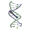
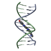
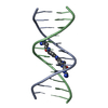
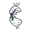
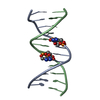
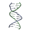
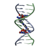
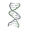
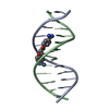
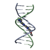
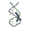
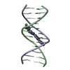
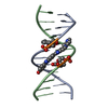
- PDB-1eel: STRUCTURE OF A COMPLEX BETWEEN THE DNA SEQUENCE DCGCGAATTCGCG AND... -
+ Open data

- Basic information
| Entry | Database: PDB / ID: 1eel | ||||||||||||||||||
|---|---|---|---|---|---|---|---|---|---|---|---|---|---|---|---|---|---|---|---|
| Title | STRUCTURE OF A COMPLEX BETWEEN THE DNA SEQUENCE DCGCGAATTCGCG AND BIS[PIPERIDINO-ETHYL]-FURAMIDINE | ||||||||||||||||||
 Components Components | 5'-D(* Keywords KeywordsDNA / MINOR GROOVE BINDING LIGAND / B-DNA / BULKY SIDE-CHAINS | Function / homology | Chem-D24 / DNA / DNA (> 10) |  Function and homology information Function and homology informationMethod |  X-RAY DIFFRACTION / MOLECULAR REPLACEMENT / Resolution: 2.4 Å X-RAY DIFFRACTION / MOLECULAR REPLACEMENT / Resolution: 2.4 Å  Authors AuthorsNeidle, S. / Simpson, I.J. |  CitationJournal: J.Mol.Biol. / Year: 2000 CitationJournal: J.Mol.Biol. / Year: 2000Title: A thermodynamic and structural analysis of DNA minor-groove complex formation. Authors: Mazur, S. / Tanious, F.A. / Ding, D. / Kumar, A. / Boykin, D.W. / Simpson, I.J. / Neidle, S. / Wilson, W.D. History |
|
- Structure visualization
| Structure viewer | Molecule: MolmilJmol/JSmol |
|---|
- Downloads & links
-Download
| PDBx/mmCIF format | 1eel.cif.gz | 25.3 KB | Display | PDBx/mmCIF format |
|---|---|---|---|---|
| PDB format | pdb1eel.ent.gz | 16 KB | Display | PDB format |
| PDBx/mmJSON format | 1eel.json.gz | Tree view | PDBx/mmJSON format | |
| Others |  Other downloads Other downloads |
-Validation report
| Summary document | 1eel_validation.pdf.gz | 390.9 KB | Display | wwPDB validaton report |
|---|---|---|---|---|
| Full document | 1eel_full_validation.pdf.gz | 393.8 KB | Display | |
| Data in XML | 1eel_validation.xml.gz | 2 KB | Display | |
| Data in CIF | 1eel_validation.cif.gz | 3.2 KB | Display | |
| Arichive directory | https://data.pdbj.org/pub/pdb/validation_reports/ee/1eelftp://data.pdbj.org/pub/pdb/validation_reports/ee/1eel | HTTPS FTP |
-Related structure data
| Similar structure data |
|---|


-Links
 PDBj
PDBj


- Assembly
| Deposited unit | 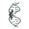
| ||||||||||
|---|---|---|---|---|---|---|---|---|---|---|---|
| 1 |
| ||||||||||
| Unit cell |
|
-Components
| #1: DNA chain | Mass: 3663.392 Da / Num. of mol.: 2 / Source method: obtained synthetically #2: Chemical | ChemComp-D24 / |   Mass: 436.548 Da / Num. of mol.: 1 / Source method: obtained synthetically / Formula: C28H28N4O Mass: 436.548 Da / Num. of mol.: 1 / Source method: obtained synthetically / Formula: C28H28N4O#3: Water | ChemComp-HOH / |  Mass: 18.015 Da / Num. of mol.: 90 / Source method: isolated from a natural source / Formula: H2O Mass: 18.015 Da / Num. of mol.: 90 / Source method: isolated from a natural source / Formula: H2O |
|---|
-Experimental details
-Experiment
| Experiment | Method: X-RAY DIFFRACTION / Number of used crystals: 1 |
|---|
- Sample preparation
| Crystal | Density Matthews: 2.37 Å3/Da / Density % sol: 48.12 % | ||||||||||||||||||||||||||||||||||||||||||
|---|---|---|---|---|---|---|---|---|---|---|---|---|---|---|---|---|---|---|---|---|---|---|---|---|---|---|---|---|---|---|---|---|---|---|---|---|---|---|---|---|---|---|---|
| Crystal grow | Temperature: 293 K / Method: vapor diffusion, hanging drop / pH: 7 Details: MPD 3 UL OF 25%, MGCL2 2UL OF 90MM, SPERMINE 1UL OF 10MM, DNA 2UL OF 2MM, LIGAND 2UL OF 3MM, MPD IN RESERVOIR 1ML OF 30%, pH 7, VAPOR DIFFUSION, HANGING DROP, temperature 293K | ||||||||||||||||||||||||||||||||||||||||||
| Components of the solutions |
| ||||||||||||||||||||||||||||||||||||||||||
| Crystal grow | *PLUS Temperature: 286 K / Method: vapor diffusion, sitting drop | ||||||||||||||||||||||||||||||||||||||||||
| Components of the solutions | *PLUS
|
-Data collection
| Diffraction | Mean temperature: 293 K |
|---|---|
| Diffraction source | Source: ROTATING ANODE / Type: RIGAKU RU200 / Wavelength: 1.5418 |
| Detector | Type: RIGAKU RAXIS IIC / Detector: IMAGE PLATE / Date: Oct 20, 1998 / Details: YALE FOCUSSING MIRRORS |
| Radiation | Monochromator: Y / Protocol: SINGLE WAVELENGTH / Monochromatic (M) / Laue (L): M / Scattering type: x-ray |
| Radiation wavelength | Wavelength: 1.5418 Å / Relative weight: 1 |
| Reflection | Resolution: 2.4→12 Å / Num. obs: 3105 / % possible obs: 94.3 % / Observed criterion σ(F): 2 / Observed criterion σ(I): 1 / Redundancy: 6 % / Rmerge(I) obs: 0.033 / Net I/σ(I): 9.6 |
| Reflection | *PLUS Highest resolution: 2.4 Å / Lowest resolution: 12 Å / Num. all: 3292 / Num. measured all: 18138 |
- Processing
| Software |
| |||||||||||||||||||||||||||||||||
|---|---|---|---|---|---|---|---|---|---|---|---|---|---|---|---|---|---|---|---|---|---|---|---|---|---|---|---|---|---|---|---|---|---|---|
| Refinement | Method to determine structure: MOLECULAR REPLACEMENT Starting model: DNA PART OF NDB STRUCTURE GDL044 Resolution: 2.4→8 Å / Cross valid method: THROUGHOUT / σ(F): 2 / σ(I): 1 / Stereochemistry target values: PARKINSON ET AL. / Details: USED SHELX-97 PROCEDURE
| |||||||||||||||||||||||||||||||||
| Refinement step | Cycle: LAST / Resolution: 2.4→8 Å
| |||||||||||||||||||||||||||||||||
| Refine LS restraints |
| |||||||||||||||||||||||||||||||||
| Software | *PLUS Name: SHELXL / Version: 97 / Classification: refinement | |||||||||||||||||||||||||||||||||
| Refinement | *PLUS Highest resolution: 2.4 Å / Rfactor all: 0.184 / Rfactor Rwork: 0.184 | |||||||||||||||||||||||||||||||||
| Solvent computation | *PLUS | |||||||||||||||||||||||||||||||||
| Displacement parameters | *PLUS |
