 Movie
Movie Controller

[English] 日本語
 Yorodumi
- PDB-3gjj: crystal structure of a DNA duplex containing 7,8-dihydropyridol[2... -
+ Open data

- Basic information
| Entry | Database: PDB / ID: 3gjj | ||||||||||||||||||
|---|---|---|---|---|---|---|---|---|---|---|---|---|---|---|---|---|---|---|---|
| Title | crystal structure of a DNA duplex containing 7,8-dihydropyridol[2,3-d]pyrimidin-2-one | ||||||||||||||||||
 Components Components | 5'-D(* Keywords KeywordsDNA / duplex / bicyclic cytosine / 7 / 8-dihydropyridol[2 / 3-d]pyrimidin-2-one | Function / homology | Chem-HT / DNA / DNA (> 10) |  Function and homology information Function and homology informationMethod |  X-RAY DIFFRACTION / SYNCHROTRON / MOLECULAR REPLACEMENT / Resolution: 2.9 Å X-RAY DIFFRACTION / SYNCHROTRON / MOLECULAR REPLACEMENT / Resolution: 2.9 Å  Authors AuthorsTakenaka, A. / Juan, E.C.M. / Shimizu, S. / Haraguchi, T. / Xiao, M. / Kurose, T. / Ohkubo, A. / Sekine, M. / Shibata, T. / Millington, C.L. / Williams, D.M. |  CitationJournal: To be Published CitationJournal: To be PublishedTitle: Insights into the stabilizing contributions of bicyclic cytosine analogues: crystal structures of DNA duplexes containing 7,8-dihydropyridol[2,3-d]pyrimidin-2-one Authors: Takenaka, A. / Juan, E.C.M. / Shimizu, S. / Haraguchi, T. / Xiao, M. / Kurose, T. / Ohkubo, A. / Sekine, M. / Shibata, T. / Millington, M. History |
|
- Structure visualization
| Structure viewer | Molecule: MolmilJmol/JSmol |
|---|
- Downloads & links
-Download
| PDBx/mmCIF format | 3gjj.cif.gz | 25.5 KB | Display | PDBx/mmCIF format |
|---|---|---|---|---|
| PDB format | pdb3gjj.ent.gz | 16.4 KB | Display | PDB format |
| PDBx/mmJSON format | 3gjj.json.gz | Tree view | PDBx/mmJSON format | |
| Others |  Other downloads Other downloads |
-Validation report
| Arichive directory | https://data.pdbj.org/pub/pdb/validation_reports/gj/3gjjftp://data.pdbj.org/pub/pdb/validation_reports/gj/3gjj | HTTPS FTP |
|---|
-Related structure data
| Related structure data | 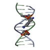 3gjhC 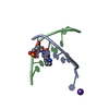 3gjkC 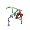 3gjlC 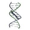 1dnhS C: citing same article ( S: Starting model for refinement |
|---|---|
| Similar structure data |


-Links
 PDBj
PDBj


- Assembly
| Deposited unit | 
| ||||||||
|---|---|---|---|---|---|---|---|---|---|
| 1 |
| ||||||||
| Unit cell |
|
-Components
| #1: DNA chain | Mass: 3686.429 Da / Num. of mol.: 2 / Source method: obtained synthetically #2: Chemical | ChemComp-HT / |   Mass: 424.498 Da / Num. of mol.: 1 / Source method: obtained synthetically / Formula: C25H24N6O Mass: 424.498 Da / Num. of mol.: 1 / Source method: obtained synthetically / Formula: C25H24N6O#3: Water | ChemComp-HOH / |  Mass: 18.015 Da / Num. of mol.: 56 / Source method: isolated from a natural source / Formula: H2O Mass: 18.015 Da / Num. of mol.: 56 / Source method: isolated from a natural source / Formula: H2O |
|---|
-Experimental details
-Experiment
| Experiment | Method: X-RAY DIFFRACTION / Number of used crystals: 1 |
|---|
- Sample preparation
| Crystal | Density Matthews: 2.28 Å3/Da / Density % sol: 46.12 % |
|---|---|
| Crystal grow | Temperature: 277 K / Method: vapor diffusion, hanging drop / pH: 6 Details: 0.60mM ssDNA, 20mM Sodium cacodylate (pH6.0), 6mM Spermine tetrahydrochloride, 40mM Potassium chloride, 6mM Sodium chloride, 0.2% CHAPS, 0.30mM Hoechst33258, 5% MPD , VAPOR DIFFUSION, ...Details: 0.60mM ssDNA, 20mM Sodium cacodylate (pH6.0), 6mM Spermine tetrahydrochloride, 40mM Potassium chloride, 6mM Sodium chloride, 0.2% CHAPS, 0.30mM Hoechst33258, 5% MPD , VAPOR DIFFUSION, HANGING DROP, temperature 277K |
-Data collection
| Diffraction | Mean temperature: 100 K |
|---|---|
| Diffraction source | Source: SYNCHROTRON / Site: Photon Factory  / Beamline: BL-6A / Wavelength: 0.978 Å / Beamline: BL-6A / Wavelength: 0.978 Å |
| Detector | Type: ADSC QUANTUM 4 / Detector: CCD / Date: Jan 26, 2008 / Details: Mirrors |
| Radiation | Protocol: SINGLE WAVELENGTH / Monochromatic (M) / Laue (L): M / Scattering type: x-ray |
| Radiation wavelength | Wavelength: 0.978 Å / Relative weight: 1 |
| Reflection | Resolution: 2.9→50 Å / Num. all: 1714 / Num. obs: 1714 / % possible obs: 99.8 % / Observed criterion σ(F): 0 / Observed criterion σ(I): 0 / Redundancy: 13.1 % / Rmerge(I) obs: 0.035 / Net I/σ(I): 69.6 |
| Reflection shell | Resolution: 2.9→3 Å / Redundancy: 13.5 % / Rmerge(I) obs: 0.298 / Mean I/σ(I) obs: 10.8 / Num. unique all: 169 / % possible all: 100 |
- Processing
| Software |
| |||||||||||||||||||||||||
|---|---|---|---|---|---|---|---|---|---|---|---|---|---|---|---|---|---|---|---|---|---|---|---|---|---|---|
| Refinement | Method to determine structure: MOLECULAR REPLACEMENT Starting model: 1DNH Resolution: 2.9→9.9 Å / Isotropic thermal model: isotropic / Cross valid method: THROUGHOUT / σ(F): 0 / σ(I): 0 / Stereochemistry target values: Engh & Huber / Details: Used weighted full matrix least squares procedure
| |||||||||||||||||||||||||
| Displacement parameters | Biso mean: 43.7 Å2
| |||||||||||||||||||||||||
| Refinement step | Cycle: LAST / Resolution: 2.9→9.9 Å
| |||||||||||||||||||||||||
| LS refinement shell | Resolution: 2.9→3.08 Å
|
