 Movie
Movie Controller

[English] 日本語
 Yorodumi
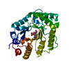
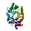
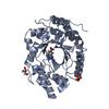
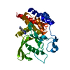
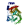
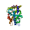
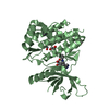
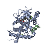
- PDB-1e0x: XYLANASE 10A FROM SREPTOMYCES LIVIDANS. XYLOBIOSYL-ENZYME INTERME... -
+ Open data

- Basic information
| Entry | Database: PDB / ID: 1e0x | |||||||||
|---|---|---|---|---|---|---|---|---|---|---|
| Title | XYLANASE 10A FROM SREPTOMYCES LIVIDANS. XYLOBIOSYL-ENZYME INTERMEDIATE AT 1.65 A | |||||||||
 Components Components | ENDO-1,4-BETA-XYLANASE A | |||||||||
 Keywords Keywords | HYDROLASE / GLYCOSIDE HYDROLASE FAMILY 10 / XYLAN DEGRADATION / GLYCOSYL-ENZYME INTERMEDIATE | |||||||||
| Function / homology |  Function and homology information Function and homology informationendo-1,4-beta-xylanase activity / endo-1,4-beta-xylanase / xylan catabolic process / carbohydrate binding / extracellular region Similarity search - Function | |||||||||
| Biological species |  STREPTOMYCES LIVIDANS (bacteria) STREPTOMYCES LIVIDANS (bacteria) | |||||||||
| Method |  X-RAY DIFFRACTION / MOLECULAR REPLACEMENT / Resolution: 1.65 Å X-RAY DIFFRACTION / MOLECULAR REPLACEMENT / Resolution: 1.65 Å | |||||||||
 Authors Authors | Ducros, V. / Charnock, S.J. / Derewenda, U. / Derewenda, Z.S. / Dauter, Z. / Dupont, C. / Shareck, F. / Morosoli, R. / Kluepfel, D. / Davies, G.J. | |||||||||
 Citation Citation | Journal: J.Biol.Chem. / Year: 2000 Title: Substrate Specificity in Glycoside Hydrolase Family 10. Structural and Kinetic Analysis of the Streptomyces Lividans Xylanase 10A Authors: Ducros, V. / Charnock, S.J. / Derewenda, U. / Derewenda, Z.S. / Dauter, Z. / Dupont, C. / Shareck, F. / Morosoli, R. / Kluepfel, D. / Davies, G.J. #1: Journal: J.Biol.Chem. / Year: 1994Title: Crystal Structure, at 2.6 A Resolution, of the Streptomyces Lividans Xylanase A, a Member of the F Family of B-1,4-D-Glycanases Authors: Derewenda, U. / Swenson, L. / Green, R. / Wei, Y. / Morosoli, R. / Shareck, F. / Kluepfel, D. / Derewenda, Z.S. | |||||||||
| History |
|
- Structure visualization
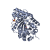
| Structure viewer | Molecule: MolmilJmol/JSmol |
|---|
- Downloads & links
-Download
| PDBx/mmCIF format | 1e0x.cif.gz | 150.1 KB | Display | PDBx/mmCIF format |
|---|---|---|---|---|
| PDB format | pdb1e0x.ent.gz | 116.4 KB | Display | PDB format |
| PDBx/mmJSON format | 1e0x.json.gz | Tree view | PDBx/mmJSON format | |
| Others |  Other downloads Other downloads |
-Validation report
| Arichive directory | https://data.pdbj.org/pub/pdb/validation_reports/e0/1e0xftp://data.pdbj.org/pub/pdb/validation_reports/e0/1e0x | HTTPS FTP |
|---|
-Related structure data


-Links
 PDBj
PDBj


- Assembly
| Deposited unit | 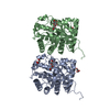
| ||||||||
|---|---|---|---|---|---|---|---|---|---|
| 1 | 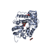
| ||||||||
| 2 | 
| ||||||||
| Unit cell |
| ||||||||
| Noncrystallographic symmetry (NCS) | NCS oper: (Code: given Matrix: (0.99908, -0.04144, 0.0114), Vector: |
-Components
| #1: Protein | Mass: 34129.457 Da / Num. of mol.: 2 / Fragment: CATALYTIC MODULE, RESIDUES 32-450 Source method: isolated from a genetically manipulated source Details: GLYCOSYL ENXYME INTERMEDIATE. COVALENT LINK BETWEEN GLU 236 AND THE SUBSTRATE Source: (gene. exp.) STREPTOMYCES LIVIDANS (bacteria) / Production host: STREPTOMYCES LIVIDANS (bacteria) / Strain (production host): IAF 19 / References: UniProt: P26514, endo-1,4-beta-xylanase#2: Polysaccharide | Source method: isolated from a genetically manipulated source #3: Chemical |   Mass: 92.094 Da / Num. of mol.: 3 / Source method: obtained synthetically / Formula: C3H8O3 Mass: 92.094 Da / Num. of mol.: 3 / Source method: obtained synthetically / Formula: C3H8O3#4: Water | ChemComp-HOH / |  Mass: 18.015 Da / Num. of mol.: 717 / Source method: isolated from a natural source / Formula: H2O Mass: 18.015 Da / Num. of mol.: 717 / Source method: isolated from a natural source / Formula: H2OHas protein modification | Y | Sequence details | THE FIRST 41 RESIDUES IN THE DATABASE CORRESPOND TO THE SIGNAL PEPTIDE. THE NUMBERING USED IN THE ...THE FIRST 41 RESIDUES IN THE DATABASE CORRESPOND | |
|---|
-Experimental details
-Experiment
| Experiment | Method: X-RAY DIFFRACTION / Number of used crystals: 1 |
|---|
- Sample preparation
| Crystal | Density Matthews: 2.7 Å3/Da / Density % sol: 44 % | |||||||||||||||||||||||||
|---|---|---|---|---|---|---|---|---|---|---|---|---|---|---|---|---|---|---|---|---|---|---|---|---|---|---|
| Crystal grow | pH: 7.5 Details: PROTEIN WAS CRYSTALLISED WITH 18 % PEG 5000 AS PRECIPITANT, 100MM HEPES PH 7.5 AS BUFFER, 10% ISOPROPANOL, CRYSTAL WERE SOAKED IN PRESENCE OF POWDERED SUBSTRATE FOR 12 HOURS. 15% GLYCEROL ...Details: PROTEIN WAS CRYSTALLISED WITH 18 % PEG 5000 AS PRECIPITANT, 100MM HEPES PH 7.5 AS BUFFER, 10% ISOPROPANOL, CRYSTAL WERE SOAKED IN PRESENCE OF POWDERED SUBSTRATE FOR 12 HOURS. 15% GLYCEROL WAS ADDED AS CRYOPROTECTANT | |||||||||||||||||||||||||
| Crystal grow | *PLUS Method: vapor diffusion, hanging drop | |||||||||||||||||||||||||
| Components of the solutions | *PLUS
|
-Data collection
| Diffraction | Mean temperature: 100 K |
|---|---|
| Diffraction source | Source: ROTATING ANODE / Type: RIGAKU RU200 / Wavelength: 1.5418 |
| Detector | Type: MARRESEARCH / Detector: IMAGE PLATE / Date: Nov 15, 1997 / Details: LONG MIRRORS (MSC) |
| Radiation | Protocol: SINGLE WAVELENGTH / Monochromatic (M) / Laue (L): M / Scattering type: x-ray |
| Radiation wavelength | Wavelength: 1.5418 Å / Relative weight: 1 |
| Reflection | Resolution: 1.65→15 Å / Num. obs: 66881 / % possible obs: 99 % / Observed criterion σ(I): 2 / Redundancy: 3.5 % / Biso Wilson estimate: 11.8 Å2 / Rmerge(I) obs: 0.033 / Net I/σ(I): 35.6 |
| Reflection shell | Resolution: 1.65→1.71 Å / Redundancy: 4.8 % / Rmerge(I) obs: 0.097 / Mean I/σ(I) obs: 12.9 / % possible all: 94 |
| Reflection | *PLUS Lowest resolution: 15 Å / % possible obs: 99 % |
| Reflection shell | *PLUS % possible obs: 94 % |
- Processing
| Software |
| ||||||||||||||||||||||||||||||||||||||||||||||||||||||||||||||||||||||||||||||||||||
|---|---|---|---|---|---|---|---|---|---|---|---|---|---|---|---|---|---|---|---|---|---|---|---|---|---|---|---|---|---|---|---|---|---|---|---|---|---|---|---|---|---|---|---|---|---|---|---|---|---|---|---|---|---|---|---|---|---|---|---|---|---|---|---|---|---|---|---|---|---|---|---|---|---|---|---|---|---|---|---|---|---|---|---|---|---|
| Refinement | Method to determine structure: MOLECULAR REPLACEMENT Starting model: NATIVE STRUCTURE AT 1.2 Resolution: 1.65→15 Å / Cross valid method: THROUGHOUT / σ(F): 0 Details: DOUBLY CONFIGURATED DISULPHIDE BOND BETWEEN CYS168 AND CYS201
| ||||||||||||||||||||||||||||||||||||||||||||||||||||||||||||||||||||||||||||||||||||
| Refinement step | Cycle: LAST / Resolution: 1.65→15 Å
| ||||||||||||||||||||||||||||||||||||||||||||||||||||||||||||||||||||||||||||||||||||
| Refine LS restraints |
| ||||||||||||||||||||||||||||||||||||||||||||||||||||||||||||||||||||||||||||||||||||
| Software | *PLUS Name: REFMAC / Classification: refinement | ||||||||||||||||||||||||||||||||||||||||||||||||||||||||||||||||||||||||||||||||||||
| Refinement | *PLUS Lowest resolution: 15 Å / Rfactor obs: 0.12 | ||||||||||||||||||||||||||||||||||||||||||||||||||||||||||||||||||||||||||||||||||||
| Solvent computation | *PLUS | ||||||||||||||||||||||||||||||||||||||||||||||||||||||||||||||||||||||||||||||||||||
| Displacement parameters | *PLUS |
