 Movie
Movie Controller

[English] 日本語
 Yorodumi
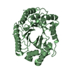
- PDB-1e0v: Xylanase 10A from Sreptomyces lividans. cellobiosyl-enzyme interm... -
+ Open data

- Basic information
| Entry | Database: PDB / ID: 1e0v | |||||||||
|---|---|---|---|---|---|---|---|---|---|---|
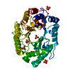
| Title | Xylanase 10A from Sreptomyces lividans. cellobiosyl-enzyme intermediate at 1.7 A | |||||||||
 Components Components | ENDO-1,4-BETA-XYLANASE A | |||||||||
 Keywords Keywords | HYDROLASE / XYLANASE / XYLAN DEGRADATION / GLYCOSYL-ENZYME INTERMEDIATE | |||||||||
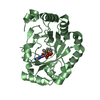
| Function / homology |  Function and homology information Function and homology informationendo-1,4-beta-xylanase / endo-1,4-beta-xylanase activity / xylan catabolic process / carbohydrate binding / extracellular region Similarity search - Function | |||||||||
| Biological species |  STREPTOMYCES LIVIDANS (bacteria) STREPTOMYCES LIVIDANS (bacteria) | |||||||||
| Method |  X-RAY DIFFRACTION / MOLECULAR REPLACEMENT / Resolution: 1.7 Å X-RAY DIFFRACTION / MOLECULAR REPLACEMENT / Resolution: 1.7 Å | |||||||||
 Authors Authors | Ducros, V. / Charnock, S.J. / Derewenda, U. / Derewenda, Z.S. / Dauter, Z. / Dupont, C. / Shareck, F. / Morosoli, R. / Kluepfel, D. / Davies, G.J. | |||||||||
 Citation Citation | Journal: J.Biol.Chem. / Year: 2000 Title: Substrate Specificity in Glycoside Hydrolase Family 10. Structural and Kinetic Analysis of the Streptomyces Lividans Xylanase 10A Authors: Ducros, V. / Charnock, S.J. / Derewenda, U. / Derewenda, Z.S. / Dauter, Z. / Dupont, C. / Shareck, F. / Morosoli, R. / Kluepfel, D. / Davies, G.J. #1: Journal: J.Biol.Chem. / Year: 2000 Title: Substrate Specificity in Glycoside Hydrolase Family 10. Tyrosine 87 and Leucine 314 Play a Pivotal Role in Discriminating between Glucose and Xylose Binding in the Proximal Active Site of ...Title: Substrate Specificity in Glycoside Hydrolase Family 10. Tyrosine 87 and Leucine 314 Play a Pivotal Role in Discriminating between Glucose and Xylose Binding in the Proximal Active Site of Pseudomonas Cellulosa Xylanase 10A. Authors: Andrews, S.R. / Charnock, S.J. / Lakey, J.H. / Davies, G.J. / Claeyssens, M. / Nerinckx, W. / Underwood, M. / Sinnott, M.L. / Warren, R.A. / Gilbert, H.J. #2: Journal: J.Biol.Chem. / Year: 1994Title: Crystal Structure, at 2.6 A Resolution, of the Streptomyces Lividans Xylanase A, a Member of the F Family of B-1,4-D-Glycanases Authors: Derewenda, U. / Swenson, L. / Green, R. / Wei, Y. / Morosoli, R. / Shareck, F. / Kluepfel, D. / Derewenda, Z.S. | |||||||||
| History |
|
- Structure visualization
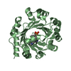
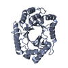
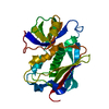
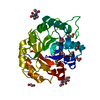
| Structure viewer | Molecule: MolmilJmol/JSmol |
|---|
- Downloads & links
-Download
| PDBx/mmCIF format | 1e0v.cif.gz | 85.1 KB | Display | PDBx/mmCIF format |
|---|---|---|---|---|
| PDB format | pdb1e0v.ent.gz | 62.4 KB | Display | PDB format |
| PDBx/mmJSON format | 1e0v.json.gz | Tree view | PDBx/mmJSON format | |
| Others |  Other downloads Other downloads |
-Validation report
| Arichive directory | https://data.pdbj.org/pub/pdb/validation_reports/e0/1e0vftp://data.pdbj.org/pub/pdb/validation_reports/e0/1e0v | HTTPS FTP |
|---|
-Related structure data


-Links
 PDBj
PDBj


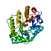
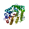
- Assembly
| Deposited unit | 
| ||||||||
|---|---|---|---|---|---|---|---|---|---|
| 1 |
| ||||||||
| Unit cell |
|
-Components
| #1: Protein | Mass: 34085.449 Da / Num. of mol.: 1 / Fragment: CATALYTIC MODULE, RESIDUES 42-343 Source method: isolated from a genetically manipulated source Details: GLYCOSYL ENXYME INTERMEDIATE. COVALENT LINK BETWEEN GLU 236 AND THE SUBSTRATE Source: (gene. exp.) STREPTOMYCES LIVIDANS (bacteria) / Production host: STREPTOMYCES LIVIDANS (bacteria) / Strain (production host): IAF 19 / References: UniProt: P26514, endo-1,4-beta-xylanase |
|---|---|
| #2: Polysaccharide | beta-D-glucopyranose-(1-4)-2-deoxy-2-fluoro-alpha-D-glucopyranose / 2-deoxy-2-fluoro-beta-cellobiose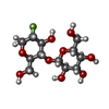  Source method: isolated from a genetically manipulated source Details: oligosaccharide / References: 2-deoxy-2-fluoro-beta-cellobiose |
| #3: Water | ChemComp-HOH /  Mass: 18.015 Da / Num. of mol.: 505 / Source method: isolated from a natural source / Formula: H2O Mass: 18.015 Da / Num. of mol.: 505 / Source method: isolated from a natural source / Formula: H2O |
| Has protein modification | Y |
| Sequence details | THE FIRST 41 RESIDUES IN THE DATABASE CORRESPOND TO THE SIGNAL PEPTIDE. THE NUMBERING USED IN THE ...THE FIRST 41 RESIDUES IN THE DATABASE CORRESPOND |
-Experimental details
-Experiment
| Experiment | Method: X-RAY DIFFRACTION / Number of used crystals: 1 |
|---|
- Sample preparation
| Crystal | Density Matthews: 2.2 Å3/Da / Density % sol: 44.3 % | |||||||||||||||||||||||||
|---|---|---|---|---|---|---|---|---|---|---|---|---|---|---|---|---|---|---|---|---|---|---|---|---|---|---|
| Crystal grow | pH: 7.5 Details: PROTEIN WAS CRYSTALLISED WITH 16 % PEG 4000 AS PRECIPITANT,100MM HEPES PH 7.5 AS BUFFER, 10% ISOPROPANOL, CRYSTAL WERE SOAKED IN PRESENCE OF POWDERED SUBSTR | |||||||||||||||||||||||||
| Crystal grow | *PLUS Method: vapor diffusion, hanging drop | |||||||||||||||||||||||||
| Components of the solutions | *PLUS
|
-Data collection
| Diffraction | Mean temperature: 100 K |
|---|---|
| Diffraction source | Source: ROTATING ANODE / Type: RIGAKU RU200 / Wavelength: 1.5418 |
| Detector | Type: MARRESEARCH / Detector: IMAGE PLATE / Date: Sep 15, 1999 |
| Radiation | Protocol: SINGLE WAVELENGTH / Monochromatic (M) / Laue (L): M / Scattering type: x-ray |
| Radiation wavelength | Wavelength: 1.5418 Å / Relative weight: 1 |
| Reflection | Resolution: 1.7→15 Å / Num. obs: 29439 / % possible obs: 97 % / Observed criterion σ(I): 2 / Redundancy: 4.3 % / Biso Wilson estimate: 17.5 Å2 / Rmerge(I) obs: 0.032 / Net I/σ(I): 36.5 |
| Reflection shell | Resolution: 1.7→1.76 Å / Redundancy: 4.9 % / Rmerge(I) obs: 0.121 / Mean I/σ(I) obs: 9.4 / % possible all: 82 |
| Reflection | *PLUS Lowest resolution: 15 Å / % possible obs: 97 % |
| Reflection shell | *PLUS % possible obs: 82 % |
- Processing
| Software |
| ||||||||||||||||||||||||||||||||||||||||||||||||||||||||||||||||||||||||||||||||||||
|---|---|---|---|---|---|---|---|---|---|---|---|---|---|---|---|---|---|---|---|---|---|---|---|---|---|---|---|---|---|---|---|---|---|---|---|---|---|---|---|---|---|---|---|---|---|---|---|---|---|---|---|---|---|---|---|---|---|---|---|---|---|---|---|---|---|---|---|---|---|---|---|---|---|---|---|---|---|---|---|---|---|---|---|---|---|
| Refinement | Method to determine structure: MOLECULAR REPLACEMENT Starting model: NATIVE STRUCTURE AT 1.2 Resolution: 1.7→15 Å / Cross valid method: THROUGHOUT / σ(F): 0 Details: DOUBLY CONFIGURATED DISULPHIDE BOND BETWEEN CYS168 AND CYS201 THE SIDE CHAIN OF GLN 88, TRP 274 AND ARG 275 ARE MISSING SINCE THEY ARE TOO DISORDERED TO BE BUILT INTO DENSITY
| ||||||||||||||||||||||||||||||||||||||||||||||||||||||||||||||||||||||||||||||||||||
| Refinement step | Cycle: LAST / Resolution: 1.7→15 Å
| ||||||||||||||||||||||||||||||||||||||||||||||||||||||||||||||||||||||||||||||||||||
| Refine LS restraints |
| ||||||||||||||||||||||||||||||||||||||||||||||||||||||||||||||||||||||||||||||||||||
| Software | *PLUS Name: REFMAC / Classification: refinement | ||||||||||||||||||||||||||||||||||||||||||||||||||||||||||||||||||||||||||||||||||||
| Refinement | *PLUS Lowest resolution: 15 Å / Rfactor obs: 0.15 | ||||||||||||||||||||||||||||||||||||||||||||||||||||||||||||||||||||||||||||||||||||
| Solvent computation | *PLUS | ||||||||||||||||||||||||||||||||||||||||||||||||||||||||||||||||||||||||||||||||||||
| Displacement parameters | *PLUS |
