 Movie
Movie Controller


+ Open data

- Basic information
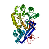
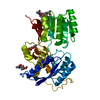
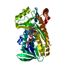
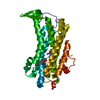
| Entry | Database: PDB / ID: 1dzg | |||||||||
|---|---|---|---|---|---|---|---|---|---|---|
| Title | N135Q-S380C-ANTITHROMBIN-III | |||||||||
 Components Components | (ANTITHROMBIN- ...) x 2 | |||||||||
 Keywords Keywords |  BLOOD CLOTTING / SERPIN BLOOD CLOTTING / SERPIN | |||||||||
| Function / homology |  Function and homology informationregulation of blood coagulation / Common Pathway of Fibrin Clot Formation / Intrinsic Pathway of Fibrin Clot Formation / Post-translational protein phosphorylation / serine-type endopeptidase inhibitor activity / Regulation of Insulin-like Growth Factor (IGF) transport and uptake by Insulin-like Growth Factor Binding Proteins (IGFBPs) / blood coagulation / heparin binding / collagen-containing extracellular matrix / blood microparticle ...regulation of blood coagulation / Common Pathway of Fibrin Clot Formation / Intrinsic Pathway of Fibrin Clot Formation / Post-translational protein phosphorylation / serine-type endopeptidase inhibitor activity / Regulation of Insulin-like Growth Factor (IGF) transport and uptake by Insulin-like Growth Factor Binding Proteins (IGFBPs) / blood coagulation / heparin binding / collagen-containing extracellular matrix / blood microparticle / protease binding / endoplasmic reticulum lumen / extracellular space / extracellular exosome / extracellular region / identical protein binding / plasma membrane Function and homology informationregulation of blood coagulation / Common Pathway of Fibrin Clot Formation / Intrinsic Pathway of Fibrin Clot Formation / Post-translational protein phosphorylation / serine-type endopeptidase inhibitor activity / Regulation of Insulin-like Growth Factor (IGF) transport and uptake by Insulin-like Growth Factor Binding Proteins (IGFBPs) / blood coagulation / heparin binding / collagen-containing extracellular matrix / blood microparticle ...regulation of blood coagulation / Common Pathway of Fibrin Clot Formation / Intrinsic Pathway of Fibrin Clot Formation / Post-translational protein phosphorylation / serine-type endopeptidase inhibitor activity / Regulation of Insulin-like Growth Factor (IGF) transport and uptake by Insulin-like Growth Factor Binding Proteins (IGFBPs) / blood coagulation / heparin binding / collagen-containing extracellular matrix / blood microparticle / protease binding / endoplasmic reticulum lumen / extracellular space / extracellular exosome / extracellular region / identical protein binding / plasma membraneSimilarity search - Function | |||||||||
| Biological species |  HOMO SAPIENS (human) HOMO SAPIENS (human) | |||||||||
| Method | X-RAY DIFFRACTION / SYNCHROTRON / MOLECULAR REPLACEMENT / Resolution: 2.8 Å | |||||||||
 Authors Authors | McCoy, A.J. / Huntington, J.A. / Carrell, R.W. | |||||||||
 Citation Citation | Journal: J.Biol.Chem. / Year: 2000 Title: The Conformational Activation of Antithrombin. A 2. 85-A Structure of a Fluorescein Derivative Reveals an Electrostatic Link between the Hinge and Heparin Binding Regions. Authors: Huntington, J.A. / Mccoy, A.J. / Belzar, K.J. / Pei, X.Y. / Gettins, P.G.W. / Carrell, R.W. | |||||||||
| History |
|
- Structure visualization
| Structure viewer | Molecule: MolmilJmol/JSmol |
|---|
- Downloads & links
-Download
| PDBx/mmCIF format | 1dzg.cif.gz | 177.8 KB | Display | PDBx/mmCIF format |
|---|---|---|---|---|
| PDB format | pdb1dzg.ent.gz | 138.9 KB | Display | PDB format |
| PDBx/mmJSON format | 1dzg.json.gz | Tree view | PDBx/mmJSON format | |
| Others |  Other downloads Other downloads |
-Validation report
| Arichive directory | https://data.pdbj.org/pub/pdb/validation_reports/dz/1dzgftp://data.pdbj.org/pub/pdb/validation_reports/dz/1dzg | HTTPS FTP |
|---|
-Related structure data
| Related structure data |  1dzhC  2antS S: Starting model for refinement C: citing same article ( |
|---|---|
| Similar structure data |


-Links
 PDBj
PDBj


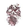
- Assembly
| Deposited unit | 
| ||||||||
|---|---|---|---|---|---|---|---|---|---|
| 1 | 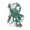
| ||||||||
| 2 | 
| ||||||||
| Unit cell |
|
-Components
-ANTITHROMBIN- ... , 2 types, 2 molecules IL
| #1: Protein | / ATIII / SERPIN C1 Mass: 49131.105 Da / Num. of mol.: 1 / Mutation: YES Source method: isolated from a genetically manipulated source Details: INHIBITORY CONFORMATION / Source: (gene. exp.) HOMO SAPIENS (human)Description: RECOMBINANT. BABY HAMSTER KIDNEY (BHK) CELL EXPRESSION Plasmid: PMASTOP / Production host:  CRICETIDAE SP. (mammal) / References: UniProt: P01008 CRICETIDAE SP. (mammal) / References: UniProt: P01008 |
|---|---|
| #2: Protein | / ATIII / SERPIN C1 Mass: 49101.016 Da / Num. of mol.: 1 / Source method: isolated from a natural source / Details: PLASMA ALPHA ANTITHROMBIN-III / Source: (natural) HOMO SAPIENS (human) / References: UniProt: P01008 |
-Sugars , 2 types, 6 molecules 
| #3: Polysaccharide | / Mass: 424.401 Da / Num. of mol.: 3 Source method: isolated from a genetically manipulated source #4: Sugar | N-Acetylglucosamine Type: D-saccharide, beta linking / Mass: 221.208 Da / Num. of mol.: 3 Type: D-saccharide, beta linking / Mass: 221.208 Da / Num. of mol.: 3Source method: isolated from a genetically manipulated source Formula: C8H15NO6 |
|---|
-Non-polymers , 2 types, 6 molecules 

| #5: Chemical | Glycerol Mass: 92.094 Da / Num. of mol.: 2 / Source method: obtained synthetically / Formula: C3H8O3 Mass: 92.094 Da / Num. of mol.: 2 / Source method: obtained synthetically / Formula: C3H8O3#6: Water | ChemComp-HOH / | WaterMass: 18.015 Da / Num. of mol.: 4 / Source method: isolated from a natural source / Formula: H2O |
|---|
-Details
| Compound details | CHAIN I: HAS ENGINEERED MUTATIONS N135Q AND S380C N135Q MUTATION TO REMOVE GLYCOSYLATION SITE S380C ...CHAIN I: HAS ENGINEERED |
|---|---|
| Sequence details | ANT3_HUMAN RESIDUE NUMBERING IN SWS IS FROM START OF ANTITHROMBIN-III LEADER SEQUENCE. STRUCTURE ...ANT3_HUMAN RESIDUE NUMBERING IN SWS IS FROM START OF ANTITHROMB |
-Experimental details
-Experiment
| Experiment | Method: X-RAY DIFFRACTION / Number of used crystals: 1 |
|---|
- Sample preparation
| Crystal | Density Matthews: 3.17 Å3/Da / Density % sol: 62 % | ||||||||||||||||||||
|---|---|---|---|---|---|---|---|---|---|---|---|---|---|---|---|---|---|---|---|---|---|
| Crystal grow | pH: 6.5 Details: 5MG/ML 1:1 MIX OF INHIBITORY: LATENT ANTITHROMBIN-III, 10.5% PEG 4000, 65 MM NACACODYLATE, PH 6.5 | ||||||||||||||||||||
| Crystal grow | *PLUS Method: batch method | ||||||||||||||||||||
| Components of the solutions | *PLUS
|
-Data collection
| Diffraction | Mean temperature: 100 K |
|---|---|
| Diffraction source | Source: SYNCHROTRON / Site: SRS  / Beamline: PX9.6 / Wavelength: 0.88 / Beamline: PX9.6 / Wavelength: 0.88 |
| Detector | Type: MARRESEARCH / Detector: CCD |
| Radiation | Protocol: SINGLE WAVELENGTH / Monochromatic (M) / Laue (L): M / Scattering type: x-ray |
| Radiation wavelength | Wavelength: 0.88 Å / Relative weight: 1 |
| Reflection | Resolution: 2.8→20 Å / Num. obs: 98714 / % possible obs: 98.3 % / Observed criterion σ(I): 0 / Redundancy: 3.5 % / Rmerge(I) obs: 0.131 / Net I/σ(I): 7.4 |
| Reflection | *PLUS Lowest resolution: 20 Å / Num. obs: 27983 / Num. measured all: 98714 |
- Processing
| Software |
| ||||||||||||||||||||||||||||||||||||||||||||||||||||||||||||||||||||||||||||||||||||
|---|---|---|---|---|---|---|---|---|---|---|---|---|---|---|---|---|---|---|---|---|---|---|---|---|---|---|---|---|---|---|---|---|---|---|---|---|---|---|---|---|---|---|---|---|---|---|---|---|---|---|---|---|---|---|---|---|---|---|---|---|---|---|---|---|---|---|---|---|---|---|---|---|---|---|---|---|---|---|---|---|---|---|---|---|---|
| Refinement | Method to determine structure: MOLECULAR REPLACEMENT Starting model: 2ANT Resolution: 2.8→20 Å / SU B: 23.25 / SU ML: 0.4309 / Cross valid method: THROUGHOUT / σ(F): 0 / ESU R: 6.82723 / ESU R Free: 0.42762 / Details: BULK SOLVENT CORRECTION CALCULATED WITH CNS
| ||||||||||||||||||||||||||||||||||||||||||||||||||||||||||||||||||||||||||||||||||||
| Displacement parameters | Biso mean: 66 Å2 | ||||||||||||||||||||||||||||||||||||||||||||||||||||||||||||||||||||||||||||||||||||
| Refinement step | Cycle: LAST / Resolution: 2.8→20 Å
| ||||||||||||||||||||||||||||||||||||||||||||||||||||||||||||||||||||||||||||||||||||
| Refine LS restraints |
| ||||||||||||||||||||||||||||||||||||||||||||||||||||||||||||||||||||||||||||||||||||
| Software | *PLUS Name: REFMAC / Classification: refinement | ||||||||||||||||||||||||||||||||||||||||||||||||||||||||||||||||||||||||||||||||||||
| Refinement | *PLUS Rfactor obs: 0.228 | ||||||||||||||||||||||||||||||||||||||||||||||||||||||||||||||||||||||||||||||||||||
| Solvent computation | *PLUS | ||||||||||||||||||||||||||||||||||||||||||||||||||||||||||||||||||||||||||||||||||||
| Displacement parameters | *PLUS | ||||||||||||||||||||||||||||||||||||||||||||||||||||||||||||||||||||||||||||||||||||
| Refine LS restraints | *PLUS Type: p_bond_d / Dev ideal: 0.011 |
