 Movie
Movie Controller

[English] 日本語
 Yorodumi
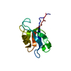
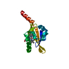
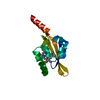
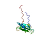
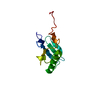
- PDB-1d5g: SOLUTION STRUCTURE OF THE PDZ2 DOMAIN FROM HUMAN PHOSPHATASE HPTP... -
+ Open data

- Basic information
| Entry | Database: PDB / ID: 1d5g | ||||||
|---|---|---|---|---|---|---|---|
| Title | SOLUTION STRUCTURE OF THE PDZ2 DOMAIN FROM HUMAN PHOSPHATASE HPTP1E COMPLEXED WITH A PEPTIDE | ||||||
 Components Components |
| ||||||
 Keywords Keywords | HYDROLASE / PROTEIN-PEPTIDE COMPLEX | ||||||
| Function / homology |  Function and homology information Function and homology informationnegative regulation of excitatory synapse assembly / regulation of phosphatidylinositol 3-kinase/protein kinase B signal transduction / phosphatidylinositol 3-kinase regulatory subunit binding / cellular response to toxic substance / Interleukin-37 signaling / RND1 GTPase cycle / RND2 GTPase cycle / RND3 GTPase cycle / protein dephosphorylation / negative regulation of protein phosphorylation ...negative regulation of excitatory synapse assembly / regulation of phosphatidylinositol 3-kinase/protein kinase B signal transduction / phosphatidylinositol 3-kinase regulatory subunit binding / cellular response to toxic substance / Interleukin-37 signaling / RND1 GTPase cycle / RND2 GTPase cycle / RND3 GTPase cycle / protein dephosphorylation / negative regulation of protein phosphorylation / Synthesis of PIPs at the plasma membrane / peptidyl-tyrosine dephosphorylation / protein-tyrosine-phosphatase / protein tyrosine phosphatase activity / lamellipodium / cell body / cytoskeleton / extracellular exosome / nucleoplasm / nucleus / plasma membrane / cytoplasm Similarity search - Function | ||||||
| Biological species |  Homo sapiens (human) Homo sapiens (human) | ||||||
| Method | SOLUTION NMR / simulated annealing | ||||||
 Authors Authors | Kozlov, G. / Gehring, K. / Ekiel, I. | ||||||
 Citation Citation | Journal: J.Mol.Biol. / Year: 2002 Title: Solution Structure of the PDZ2 Domain from Cytosolic Human Phosphatase hPTP1E Complexed with a Peptide Reveals Contribution of the beta2-beta3 Loop to PDZ Domain-Ligand Interactions Authors: Kozlov, G. / Banville, D. / Gehring, K. / Ekiel, I. #1: Journal: Biochemistry / Year: 2000Title: Solution Structure of the PDZ2 Domain from Human Phosphatase hPTP1E and its Interactions with C-terminal Peptides from the Fas Receptor Authors: Kozlov, G. / Gehring, K. / Ekiel, I. | ||||||
| History |
|
- Structure visualization
| Structure viewer | Molecule: MolmilJmol/JSmol |
|---|
- Downloads & links
-Download
| PDBx/mmCIF format | 1d5g.cif.gz | 632.3 KB | Display | PDBx/mmCIF format |
|---|---|---|---|---|
| PDB format | pdb1d5g.ent.gz | 530.3 KB | Display | PDB format |
| PDBx/mmJSON format | 1d5g.json.gz | Tree view | PDBx/mmJSON format | |
| Others |  Other downloads Other downloads |
-Validation report
| Arichive directory | https://data.pdbj.org/pub/pdb/validation_reports/d5/1d5gftp://data.pdbj.org/pub/pdb/validation_reports/d5/1d5g | HTTPS FTP |
|---|
-Related structure data
| Related structure data | |
|---|---|
| Similar structure data |


-Links
 PDBj
PDBj


- Assembly
| Deposited unit | 
| |||||||||
|---|---|---|---|---|---|---|---|---|---|---|
| 1 |
| |||||||||
| NMR ensembles |
|
-Components
| #1: Protein | Mass: 10020.252 Da / Num. of mol.: 1 / Fragment: PDZ2 DOMAIN Source method: isolated from a genetically manipulated source Source: (gene. exp.) Homo sapiens (human) / Plasmid: PET / Production host:  |
|---|---|
| #2: Protein/peptide | Mass: 1610.588 Da / Num. of mol.: 1 / Source method: obtained synthetically / Details: THE PEPTIDE WAS CHEMICALLY SYNTHESIZED. |
-Experimental details
-Experiment
| Experiment | Method: SOLUTION NMR | ||||||||||||||||||||
|---|---|---|---|---|---|---|---|---|---|---|---|---|---|---|---|---|---|---|---|---|---|
| NMR experiment |
| ||||||||||||||||||||
| NMR details | Text: THE STRUCTURE WAS DETERMINED USING TRIPLE-RESONANCE NMR SPECTROSCOPY AND 2D HOMONUCLEAR TECHNIQUES |
- Sample preparation
| Details |
| |||||||||||||||||||||||||
|---|---|---|---|---|---|---|---|---|---|---|---|---|---|---|---|---|---|---|---|---|---|---|---|---|---|---|
| Sample conditions |
| |||||||||||||||||||||||||
| Crystal grow | *PLUS Method: other / Details: NMR |
-NMR measurement
| NMR spectrometer | Type: Bruker DRX / Manufacturer: Bruker / Model: DRX / Field strength: 500 MHz |
|---|
- Processing
| NMR software |
| ||||||||||||||||||||||||||||
|---|---|---|---|---|---|---|---|---|---|---|---|---|---|---|---|---|---|---|---|---|---|---|---|---|---|---|---|---|---|
| Refinement | Method: simulated annealing / Software ordinal: 1 Details: THE STRUCTURE IS BASED ON A TOTAL OF 1391 NON-REDUNDANT CONSTRAINTS, 1269 ARE NOE-DERIVED DISTANCE CONSTRAINTS, 81 DIHEDRAL ANGLE RESTRAINTS, 41 HYDROGEN BOND CONSTRAINTS. | ||||||||||||||||||||||||||||
| NMR representative | Selection criteria: lowest energy | ||||||||||||||||||||||||||||
| NMR ensemble | Conformer selection criteria: structures with the lowest energy Conformers calculated total number: 100 / Conformers submitted total number: 20 |
