 Movie
Movie Controller

[English] 日本語
 Yorodumi
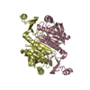
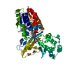
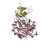
- PDB-1d02: CRYSTAL STRUCTURE OF MUNI RESTRICTION ENDONUCLEASE IN COMPLEX WIT... -
+ Open data

- Basic information
| Entry | Database: PDB / ID: 1d02 | ||||||
|---|---|---|---|---|---|---|---|
| Title | CRYSTAL STRUCTURE OF MUNI RESTRICTION ENDONUCLEASE IN COMPLEX WITH COGNATE DNA | ||||||
 Components Components |
| ||||||
 Keywords Keywords | HYDROLASE/DNA / ALPHA/BETA PROTEIN /  PROTEIN-DNA COMPLEX / DISTORTED DOUBLE HELIX / HYDROLASE-DNA COMPLEX PROTEIN-DNA COMPLEX / DISTORTED DOUBLE HELIX / HYDROLASE-DNA COMPLEX | ||||||
| Function / homology |  Function and homology informationtype II site-specific deoxyribonuclease / type II site-specific deoxyribonuclease activity / DNA restriction-modification system / DNA binding Function and homology informationtype II site-specific deoxyribonuclease / type II site-specific deoxyribonuclease activity / DNA restriction-modification system / DNA bindingSimilarity search - Function | ||||||
| Biological species |  Mycoplasma (bacteria) Mycoplasma (bacteria) | ||||||
| Method | X-RAY DIFFRACTION / SYNCHROTRON / Resolution: 1.7 Å | ||||||
 Authors Authors | Deibert, M. / Grazulis, S. / Janulaitis, A. / Siksnys, V. / Huber, R. | ||||||
 Citation Citation | Journal: EMBO J. / Year: 1999 Title: Crystal structure of MunI restriction endonuclease in complex with cognate DNA at 1.7 A resolution. Authors: Deibert, M. / Grazulis, S. / Janulaitis, A. / Siksnys, V. / Huber, R. | ||||||
| History |
|
- Structure visualization
| Structure viewer | Molecule: MolmilJmol/JSmol |
|---|
- Downloads & links
-Download
| PDBx/mmCIF format | 1d02.cif.gz | 116.2 KB | Display | PDBx/mmCIF format |
|---|---|---|---|---|
| PDB format | pdb1d02.ent.gz | 87.1 KB | Display | PDB format |
| PDBx/mmJSON format | 1d02.json.gz | Tree view | PDBx/mmJSON format | |
| Others |  Other downloads Other downloads |
-Validation report
| Arichive directory | https://data.pdbj.org/pub/pdb/validation_reports/d0/1d02ftp://data.pdbj.org/pub/pdb/validation_reports/d0/1d02 | HTTPS FTP |
|---|
-Related structure data
| Similar structure data |
|---|


-Links
 PDBj
PDBj


- Assembly
| Deposited unit | 
| ||||||||
|---|---|---|---|---|---|---|---|---|---|
| 1 |
| ||||||||
| Unit cell |
|
-Components
| #1: DNA chain | Mass: 3045.005 Da / Num. of mol.: 2 / Source method: obtained synthetically Details: DECAMERIC OLIGONUCLEOTIDE 5'-DS(GPCPCPAPAPTPTPGPGPC) WAS SYNTHESIZED WITH STANDARD PROCEDURES #2: Protein | Mass: 23376.451 Da / Num. of mol.: 2 / Mutation: D83A Source method: isolated from a genetically manipulated source Source: (gene. exp.) Mycoplasma (bacteria) / Genus: Mycoplasma / Production host: Escherichia coli (E. coli) / Strain (production host): ER2267References: UniProt: P43642, type II site-specific deoxyribonuclease#3: Water | ChemComp-HOH / | Water Mass: 18.015 Da / Num. of mol.: 646 / Source method: isolated from a natural source / Formula: H2O Mass: 18.015 Da / Num. of mol.: 646 / Source method: isolated from a natural source / Formula: H2O |
|---|
-Experimental details
-Experiment
| Experiment | Method: X-RAY DIFFRACTION / Number of used crystals: 1 |
|---|
- Sample preparation
| Crystal | Density Matthews: 2.42 Å3/Da / Density % sol: 49.25 % | ||||||||||||||||||||||||||||||||||||
|---|---|---|---|---|---|---|---|---|---|---|---|---|---|---|---|---|---|---|---|---|---|---|---|---|---|---|---|---|---|---|---|---|---|---|---|---|---|
| Crystal grow | Temperature: 293 K / Method: vapor diffusion, sitting drop / pH: 6 Details: PEG 8000, CALCIUM CHLORIDE, SODIUM CHLORIDE, MES, pH 6.0, VAPOR DIFFUSION, SITTING DROP, temperature 293.0K | ||||||||||||||||||||||||||||||||||||
| Crystal | *PLUS Density % sol: 54 % | ||||||||||||||||||||||||||||||||||||
| Crystal grow | *PLUS Temperature: 20 ℃ | ||||||||||||||||||||||||||||||||||||
| Components of the solutions | *PLUS
|
-Data collection
| Diffraction | Mean temperature: 77 K |
|---|---|
| Diffraction source | Source: SYNCHROTRON / Site: MPG/DESY, HAMBURG  / Beamline: BW6 / Wavelength: 1.006 / Beamline: BW6 / Wavelength: 1.006 |
| Detector | Type: MARRESEARCH / Detector: IMAGE PLATE |
| Radiation | Protocol: SINGLE WAVELENGTH / Monochromatic (M) / Laue (L): M / Scattering type: x-ray |
| Radiation wavelength | Wavelength: 1.006 Å / Relative weight: 1 |
| Reflection | Resolution: 1.7→8 Å / Num. all: 177834 / Num. obs: 56945 / % possible obs: 93.6 % / Observed criterion σ(I): 2 / Redundancy: 3.1 % / Rmerge(I) obs: 0.052 |
| Reflection | *PLUS Num. measured all: 177834 |
| Reflection shell | *PLUS % possible obs: 84 % |
- Processing
| Software |
| ||||||||||||||||||||||||||||||||||||||||||||||||||||||||||||
|---|---|---|---|---|---|---|---|---|---|---|---|---|---|---|---|---|---|---|---|---|---|---|---|---|---|---|---|---|---|---|---|---|---|---|---|---|---|---|---|---|---|---|---|---|---|---|---|---|---|---|---|---|---|---|---|---|---|---|---|---|---|
| Refinement | Resolution: 1.7→8 Å / σ(F): 2 Stereochemistry target values: ENGH & HUBER (PROTEIN), PARKINSON (DNA)
| ||||||||||||||||||||||||||||||||||||||||||||||||||||||||||||
| Refinement step | Cycle: LAST / Resolution: 1.7→8 Å
| ||||||||||||||||||||||||||||||||||||||||||||||||||||||||||||
| Refine LS restraints |
| ||||||||||||||||||||||||||||||||||||||||||||||||||||||||||||
| Software | *PLUS Name: CNS / Classification: refinement | ||||||||||||||||||||||||||||||||||||||||||||||||||||||||||||
| Refinement | *PLUS Highest resolution: 1.7 Å / Lowest resolution: 8 Å / σ(F): 2 / % reflection Rfree: 5 % / Rfactor obs: 0.18 / Rfactor Rwork: 0.18 | ||||||||||||||||||||||||||||||||||||||||||||||||||||||||||||
| Solvent computation | *PLUS | ||||||||||||||||||||||||||||||||||||||||||||||||||||||||||||
| Displacement parameters | *PLUS |
