 Movie
Movie Controller

[English] 日本語
 Yorodumi
- PDB-1c4z: STRUCTURE OF AN E6AP-UBCH7 COMPLEX: INSIGHTS INTO THE UBIQUITINAT... -
+ Open data

- Basic information
| Entry | Database: PDB / ID: 1c4z | ||||||
|---|---|---|---|---|---|---|---|
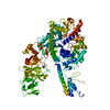
| Title | STRUCTURE OF AN E6AP-UBCH7 COMPLEX: INSIGHTS INTO THE UBIQUITINATION PATHWAY | ||||||
 Components Components |
| ||||||
 Keywords Keywords |  LIGASE / BILOBAL STRUCTURE / ELONGATED SHAPE / E3 UBIQUITIN LIGASE / E2 UBIQUITIN CONJUGATING ENZYME LIGASE / BILOBAL STRUCTURE / ELONGATED SHAPE / E3 UBIQUITIN LIGASE / E2 UBIQUITIN CONJUGATING ENZYME | ||||||
| Function / homology |  Function and homology information Function and homology informationsperm entry / positive regulation of Golgi lumen acidification / motor learning / negative regulation of dendritic spine morphogenesis / regulation of ubiquitin-dependent protein catabolic process / cell cycle phase transition / ubiquitin-protein transferase activator activity / prostate gland growth / HECT-type E3 ubiquitin transferase / protein K11-linked ubiquitination ...sperm entry / positive regulation of Golgi lumen acidification / motor learning / negative regulation of dendritic spine morphogenesis / regulation of ubiquitin-dependent protein catabolic process / cell cycle phase transition / ubiquitin-protein transferase activator activity / prostate gland growth / HECT-type E3 ubiquitin transferase / protein K11-linked ubiquitination / cellular response to glucocorticoid stimulus / positive regulation of ubiquitin-protein transferase activity / positive regulation of protein targeting to mitochondrion / E2 ubiquitin-conjugating enzyme / cellular response to steroid hormone stimulus / locomotory exploration behavior / androgen receptor signaling pathway / ubiquitin conjugating enzyme activity / progesterone receptor signaling pathway / protein K48-linked ubiquitination / protein autoubiquitination / ubiquitin ligase complex / ovarian follicle development / cellular response to brain-derived neurotrophic factor stimulus / negative regulation of TORC1 signaling / Synthesis of active ubiquitin: roles of E1 and E2 enzymes / proteasome complex / response to cocaine / response to progesterone / positive regulation of protein ubiquitination / Regulation of TNFR1 signaling / brain development / regulation of synaptic plasticity / protein modification process / response to hydrogen peroxide / regulation of circadian rhythm / Regulation of necroptotic cell death / protein polyubiquitination / ubiquitin-protein transferase activity / rhythmic process / ubiquitin protein ligase activity / Antigen processing: Ubiquitination & Proteasome degradation / E3 ubiquitin ligases ubiquitinate target proteins / ubiquitin-dependent protein catabolic process / proteasome-mediated ubiquitin-dependent protein catabolic process / cell population proliferation / transcription coactivator activity / positive regulation of phosphatidylinositol 3-kinase/protein kinase B signal transduction / protein ubiquitination / glutamatergic synapse / ubiquitin protein ligase binding / regulation of DNA-templated transcription / enzyme binding / positive regulation of transcription by RNA polymerase II / proteolysis / RNA binding / nucleoplasm / ATP binding / metal ion binding / nucleus / cytosol / cytoplasmSimilarity search - Function | ||||||
| Biological species |  Homo sapiens (human) Homo sapiens (human) | ||||||
| Method | X-RAY DIFFRACTION / SYNCHROTRON / Resolution: 2.6 Å | ||||||
 Authors Authors | Huang, L. / Kinnucan, E. / Wang, G. / Beaudenon, S. / Howley, P.M. / Huibregtse, J.M. / Pavletich, N.P. | ||||||
 Citation Citation | Journal: Science / Year: 1999 Title: Structure of an E6AP-UbcH7 complex: insights into ubiquitination by the E2-E3 enzyme cascade. Authors: Huang, L. / Kinnucan, E. / Wang, G. / Beaudenon, S. / Howley, P.M. / Huibregtse, J.M. / Pavletich, N.P. | ||||||
| History |
|
- Structure visualization
| Structure viewer | Molecule: MolmilJmol/JSmol |
|---|
- Downloads & links
-Download
| PDBx/mmCIF format | 1c4z.cif.gz | 254.2 KB | Display | PDBx/mmCIF format |
|---|---|---|---|---|
| PDB format | pdb1c4z.ent.gz | 206.2 KB | Display | PDB format |
| PDBx/mmJSON format | 1c4z.json.gz | Tree view | PDBx/mmJSON format | |
| Others |  Other downloads Other downloads |
-Validation report
| Arichive directory | https://data.pdbj.org/pub/pdb/validation_reports/c4/1c4zftp://data.pdbj.org/pub/pdb/validation_reports/c4/1c4z | HTTPS FTP |
|---|
-Related structure data


-Links
 PDBj
PDBj


- Assembly
| Deposited unit | 
| ||||||||
|---|---|---|---|---|---|---|---|---|---|
| 1 |
| ||||||||
| Unit cell |
|
-Components
| #1: Protein | Mass: 41540.434 Da / Num. of mol.: 3 / Fragment: HECT CATALYTIC DOMAIN Source method: isolated from a genetically manipulated source Source: (gene. exp.) Homo sapiens (human) / Plasmid: PGEX4T-3 / Production host:  Escherichia coli (E. coli) Escherichia coli (E. coli)References: UniProt: Q05086, Ligases; Forming carbon-nitrogen bonds; Acid-amino-acid ligases (peptide synthases)#2: Protein | | Mass: 17889.588 Da / Num. of mol.: 1 Source method: isolated from a genetically manipulated source Source: (gene. exp.) Homo sapiens (human) / Plasmid: PET / Production host: Escherichia coli (E. coli) / References: UniProt: P68036, ubiquitin-protein ligase#3: Water | ChemComp-HOH / | Water Mass: 18.015 Da / Num. of mol.: 359 / Source method: isolated from a natural source / Formula: H2O Mass: 18.015 Da / Num. of mol.: 359 / Source method: isolated from a natural source / Formula: H2O |
|---|
-Experimental details
-Experiment
| Experiment | Method: X-RAY DIFFRACTION / Number of used crystals: 1 |
|---|
- Sample preparation
| Crystal | Density Matthews: 2.53 Å3/Da / Density % sol: 51.38 % | |||||||||||||||||||||||||||||||||||
|---|---|---|---|---|---|---|---|---|---|---|---|---|---|---|---|---|---|---|---|---|---|---|---|---|---|---|---|---|---|---|---|---|---|---|---|---|
| Crystal grow | Temperature: 277 K / Method: vapor diffusion, hanging drop / pH: 6.5 Details: PEG1500, MAGNESIUM CHLORIDE, MES-NA, pH 6.5, VAPOR DIFFUSION, HANGING DROP, temperature 277.0K | |||||||||||||||||||||||||||||||||||
| Crystal grow | *PLUS | |||||||||||||||||||||||||||||||||||
| Components of the solutions | *PLUS
|
-Data collection
| Diffraction | Mean temperature: 110 K |
|---|---|
| Diffraction source | Source: SYNCHROTRON / Site: ALS  / Beamline: 5.0.2 / Wavelength: 1 / Beamline: 5.0.2 / Wavelength: 1 |
| Detector | Type: ADSC / Detector: CCD / Date: Sep 3, 1999 |
| Radiation | Protocol: SINGLE WAVELENGTH / Monochromatic (M) / Laue (L): M / Scattering type: x-ray |
| Radiation wavelength | Wavelength: 1 Å / Relative weight: 1 |
| Reflection | Resolution: 2.6→20 Å / Num. all: 106238 / Num. obs: 42425 / % possible obs: 95.8 % / Observed criterion σ(F): 0 / Observed criterion σ(I): 0 / Redundancy: 2.5 % / Biso Wilson estimate: 54 Å2 / Rmerge(I) obs: 0.055 / Net I/σ(I): 17 |
| Reflection shell | Resolution: 2.6→2.69 Å / Redundancy: 2.33 % / Rmerge(I) obs: 0.413 / % possible all: 89.5 |
| Reflection | *PLUS % possible obs: 89.8 % / Num. measured all: 106238 |
- Processing
| Software |
| ||||||||||||||||||||||||||||||||||||||||||||||||||||||||||||
|---|---|---|---|---|---|---|---|---|---|---|---|---|---|---|---|---|---|---|---|---|---|---|---|---|---|---|---|---|---|---|---|---|---|---|---|---|---|---|---|---|---|---|---|---|---|---|---|---|---|---|---|---|---|---|---|---|---|---|---|---|---|
| Refinement | Resolution: 2.6→15 Å / σ(F): 2 / σ(I): 2 / Stereochemistry target values: ENGH & HUBER (CNS LIBRARY)
| ||||||||||||||||||||||||||||||||||||||||||||||||||||||||||||
| Refinement step | Cycle: LAST / Resolution: 2.6→15 Å
| ||||||||||||||||||||||||||||||||||||||||||||||||||||||||||||
| Refine LS restraints |
| ||||||||||||||||||||||||||||||||||||||||||||||||||||||||||||
| Software | *PLUS Name: CNS / Classification: refinement | ||||||||||||||||||||||||||||||||||||||||||||||||||||||||||||
| Refinement | *PLUS Highest resolution: 2.6 Å / Lowest resolution: 15 Å / σ(F): 2 / % reflection Rfree: 5 % / Rfactor Rfree: 0.29 | ||||||||||||||||||||||||||||||||||||||||||||||||||||||||||||
| Solvent computation | *PLUS | ||||||||||||||||||||||||||||||||||||||||||||||||||||||||||||
| Displacement parameters | *PLUS | ||||||||||||||||||||||||||||||||||||||||||||||||||||||||||||
| Refine LS restraints | *PLUS Type: c_bond_d / Dev ideal: 0.011 |
