Monochromator: NI FILTER / Protocol: SINGLE WAVELENGTH / Monochromatic (M) / Laue (L): M / Scattering type: x-ray
Radiation wavelength
Wavelength: 1.5418 Å / Relative weight: 1
Reflection
Resolution: 2→20 Å / Num. obs: 16664 / % possible obs: 99.6 % / Redundancy: 5.2 % / Rmerge(I) obs: 0.063 / Rsym value: 6.3 / Net I/σ(I): 10.6
Reflection shell
Resolution: 2→2.07 Å / Rmerge(I) obs: 0.285 / Rsym value: 28.5 / % possible all: 99.4
Reflection
*PLUS
Num. measured all: 86716
Reflection shell
*PLUS
% possible obs: 99.4 %
-
Processing
Software
Name
Version
Classification
X-PLOR
3.1
refinement
DENZO
datareduction
SCALEPACK
datascaling
Refinement
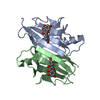
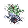
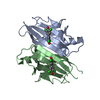
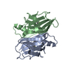
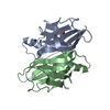
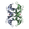
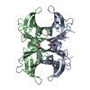
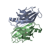
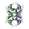
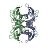
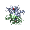
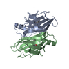
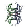
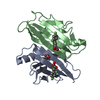
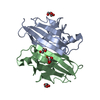
Method to determine structure: OTHER / Resolution: 2→8 Å / Rfactor Rfree error: 0.006 / Cross valid method: THROUGHOUT / σ(F): 3 Details: THE STRUCTURE CONTAINS THE FLUFENAMIC ACID MOLECULES THAT BIND IN TWO OVERLAPPING BINDING MODES IN EACH OF TWO INDEPENDENT BINDING SITES OF THE TETRAMER. SINCE THE BINDING IS ALONG THE 2- ...Details: THE STRUCTURE CONTAINS THE FLUFENAMIC ACID MOLECULES THAT BIND IN TWO OVERLAPPING BINDING MODES IN EACH OF TWO INDEPENDENT BINDING SITES OF THE TETRAMER. SINCE THE BINDING IS ALONG THE 2-FOLD CRYSTALLOGRAPHIC AXIS, AN OCCUPANCY OF 0.25 CORRESPONDS TO SATURATION OF EACH OF THE BINDING SITES.
Rfactor
Num. reflection
% reflection
Selection details
Rfree
0.251
1528
10 %
RANDOM
Rwork
0.189
-
-
-
obs
0.189
15239
92.9 %
-
Displacement parameters
Biso mean: 29.7 Å2
Refinement step
Cycle: LAST / Resolution: 2→8 Å
Protein
Nucleic acid
Ligand
Solvent
Total
Num. atoms
1762
0
80
54
1896
Refine LS restraints
Refine-ID
Type
Dev ideal
X-RAY DIFFRACTION
x_bond_d
0.016
X-RAY DIFFRACTION
x_bond_d_na
X-RAY DIFFRACTION
x_bond_d_prot
X-RAY DIFFRACTION
x_angle_d
X-RAY DIFFRACTION
x_angle_d_na
X-RAY DIFFRACTION
x_angle_d_prot
X-RAY DIFFRACTION
x_angle_deg
2.037
X-RAY DIFFRACTION
x_angle_deg_na
X-RAY DIFFRACTION
x_angle_deg_prot
X-RAY DIFFRACTION
x_dihedral_angle_d
X-RAY DIFFRACTION
x_dihedral_angle_d_na
X-RAY DIFFRACTION
x_dihedral_angle_d_prot
X-RAY DIFFRACTION
x_improper_angle_d
X-RAY DIFFRACTION
x_improper_angle_d_na
X-RAY DIFFRACTION
x_improper_angle_d_prot
X-RAY DIFFRACTION
x_mcbond_it
X-RAY DIFFRACTION
x_mcangle_it
X-RAY DIFFRACTION
x_scbond_it
X-RAY DIFFRACTION
x_scangle_it
Refine LS restraints NCS
NCS model details: RESTRAINTS / Rms dev position: 0.1122 Å / Weight position: 100
LS refinement shell
Resolution: 2→2.03 Å / Total num. of bins used: 20
Rfactor
% reflection
Rfree
0.248
10 %
Rwork
0.266
-
obs
-
85.75 %
Xplor file
Refine-ID
Serial no
Param file
Topol file
X-RAY DIFFRACTION
1
PARHCSDX.PRO
TOPHCSDX.PRO
X-RAY DIFFRACTION
2
FLU_MOPAC.PAR
FLU_MOPAC.TOP
+
About Yorodumi
-
News
-
Feb 9, 2022. New format data for meta-information of EMDB entries
New format data for meta-information of EMDB entries
Version 3 of the EMDB header file is now the official format.
The previous official version 1.9 will be removed from the archive.
In the structure databanks used in Yorodumi, some data are registered as the other names, "COVID-19 virus" and "2019-nCoV". Here are the details of the virus and the list of structure data.
Jan 31, 2019. EMDB accession codes are about to change! (news from PDBe EMDB page)
EMDB accession codes are about to change! (news from PDBe EMDB page)
The allocation of 4 digits for EMDB accession codes will soon come to an end. Whilst these codes will remain in use, new EMDB accession codes will include an additional digit and will expand incrementally as the available range of codes is exhausted. The current 4-digit format prefixed with “EMD-” (i.e. EMD-XXXX) will advance to a 5-digit format (i.e. EMD-XXXXX), and so on. It is currently estimated that the 4-digit codes will be depleted around Spring 2019, at which point the 5-digit format will come into force.
The EM Navigator/Yorodumi systems omit the EMD- prefix.
Related info.:Q: What is EMD? / ID/Accession-code notation in Yorodumi/EM Navigator
Yorodumi is a browser for structure data from EMDB, PDB, SASBDB, etc.
This page is also the successor to EM Navigator detail page, and also detail information page/front-end page for Omokage search.
The word "yorodu" (or yorozu) is an old Japanese word meaning "ten thousand". "mi" (miru) is to see.
Related info.:EMDB / PDB / SASBDB / Comparison of 3 databanks / Yorodumi Search / Aug 31, 2016. New EM Navigator & Yorodumi / Yorodumi Papers / Jmol/JSmol / Function and homology information / Changes in new EM Navigator and Yorodumi
 Movie
Movie Controller

 Yorodumi
 Open data
 Basic information
 Components
 Keywords
 Function and homology information
 Homo sapiens (human)
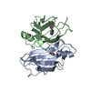
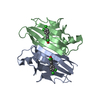
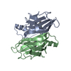
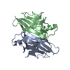
 X-RAY DIFFRACTION / OTHER / Resolution: 2 Å
  Authors
 Citation
 Structure visualization
 Downloads & links
 Other downloads

 PDBj
PDBj


 Assembly
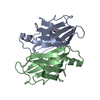


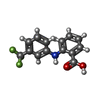
 Mass: 281.230 Da / Num. of mol.: 2 / Source method: obtained synthetically / Formula: C14H10F3NO2 / Comment: antiinflammatory, inhibitor*YM
 Mass: 18.015 Da / Num. of mol.: 54 / Source method: isolated from a natural source / Formula: H2O
 Sample preparation
 Processing