 Movie
Movie Controller

[English] 日本語
 Yorodumi
- PDB-1bll: X-RAY CRYSTALLOGRAPHIC DETERMINATION OF THE STRUCTURE OF BOVINE L... -
+ Open data

- Basic information
| Entry | Database: PDB / ID: 1bll | |||||||||
|---|---|---|---|---|---|---|---|---|---|---|
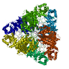
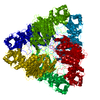
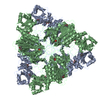
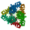
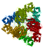
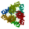
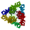
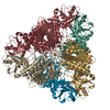
| Title | X-RAY CRYSTALLOGRAPHIC DETERMINATION OF THE STRUCTURE OF BOVINE LENS LEUCINE AMINOPEPTIDASE COMPLEXED WITH AMASTATIN: FORMULATION OF A CATALYTIC MECHANISM FEATURING A GEM-DIOLATE TRANSITION STATE | |||||||||
 Components Components |
| |||||||||
 Keywords Keywords | HYDROLASE/HYDROLASE INHIBITOR / ALPHA-AMINOACYLPEPTIDE / HYDROLASE-HYDROLASE INHIBITOR complex | |||||||||
| Function / homology |  Function and homology information Function and homology informationcysteinylglycine-S-conjugate dipeptidase / prolyl aminopeptidase / leucyl aminopeptidase / dipeptidase activity / metalloaminopeptidase activity / carboxypeptidase activity / disordered domain specific binding / peptidase activity / manganese ion binding / mitochondrion ...cysteinylglycine-S-conjugate dipeptidase / prolyl aminopeptidase / leucyl aminopeptidase / dipeptidase activity / metalloaminopeptidase activity / carboxypeptidase activity / disordered domain specific binding / peptidase activity / manganese ion binding / mitochondrion / proteolysis / cytoplasm Similarity search - Function | |||||||||
| Biological species |   Streptomyces sp. ME98-M3 (bacteria) Streptomyces sp. ME98-M3 (bacteria) | |||||||||
| Method |  X-RAY DIFFRACTION / Resolution: 2.4 Å X-RAY DIFFRACTION / Resolution: 2.4 Å | |||||||||
 Authors Authors | Kim, H. / Lipscomb, W.N. | |||||||||
 Citation Citation | Journal: Biochemistry / Year: 1993 Title: X-ray crystallographic determination of the structure of bovine lens leucine aminopeptidase complexed with amastatin: formulation of a catalytic mechanism featuring a gem-diolate transition state. Authors: Kim, H. / Lipscomb, W.N. #1: Journal: J.Mol.Biol. / Year: 1992Title: Structure Determination and Refinement of Bovine Lens Leucine Aminopeptidase and its Complex with Bestatin Authors: Burley, S.K. / David, P.R. / Sweet, R.M. / Taylor, A. / Lipscomb, W.N. #2: Journal: Proc.Natl.Acad.Sci.USA / Year: 1991Title: Leucine Aminopeptidase: Bestatin Inhibition and a Model for Enzyme-Catalyzed Peptide Hydrolysis Authors: Burley, S.K. / David, P.R. / Lipscomb, W.N. #3: Journal: Proc.Natl.Acad.Sci.USA / Year: 1990Title: Molecular Structure of Leucine Aminopeptidase at 2.7 Angstroms Resolution Authors: Burley, S.K. / David, P.R. / Taylor, A. / Lipscomb, W.N. | |||||||||
| History |
|
- Structure visualization
| Structure viewer | Molecule: MolmilJmol/JSmol |
|---|
- Downloads & links
-Download
| PDBx/mmCIF format | 1bll.cif.gz | 108.8 KB | Display | PDBx/mmCIF format |
|---|---|---|---|---|
| PDB format | pdb1bll.ent.gz | 82.8 KB | Display | PDB format |
| PDBx/mmJSON format | 1bll.json.gz | Tree view | PDBx/mmJSON format | |
| Others |  Other downloads Other downloads |
-Validation report
| Arichive directory | https://data.pdbj.org/pub/pdb/validation_reports/bl/1bllftp://data.pdbj.org/pub/pdb/validation_reports/bl/1bll | HTTPS FTP |
|---|
-Related structure data
| Similar structure data |
|---|

-Links
 PDBj
PDBj- Assembly
| Deposited unit | 
| ||||||||
|---|---|---|---|---|---|---|---|---|---|
| 1 | x 6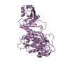
| ||||||||
| Unit cell |
| ||||||||
| Atom site foot note | 1: CIS PROLINE - PRO E 471 |
-Components
| #1: Protein | Mass: 52968.133 Da / Num. of mol.: 1 Source method: isolated from a genetically manipulated source Source: (gene. exp.) | ||||||||||
|---|---|---|---|---|---|---|---|---|---|---|---|
| #2: Protein/peptide | 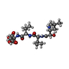  Type: Peptide-like / Class: Inhibitor / Mass: 474.548 Da / Num. of mol.: 1 / Source method: obtained synthetically / Source: (synth.) Streptomyces sp. ME98-M3 (bacteria) / References: Amastatin Type: Peptide-like / Class: Inhibitor / Mass: 474.548 Da / Num. of mol.: 1 / Source method: obtained synthetically / Source: (synth.) Streptomyces sp. ME98-M3 (bacteria) / References: Amastatin | ||||||||||
| #3: Chemical |   Mass: 65.409 Da / Num. of mol.: 2 / Source method: obtained synthetically / Formula: Zn Mass: 65.409 Da / Num. of mol.: 2 / Source method: obtained synthetically / Formula: Zn#4: Water | ChemComp-HOH / |  Mass: 18.015 Da / Num. of mol.: 132 / Source method: isolated from a natural source / Formula: H2O Mass: 18.015 Da / Num. of mol.: 132 / Source method: isolated from a natural source / Formula: H2OCompound details | THE AMASTATIN INHIBITOR, (2S,3R)3-AMINO-2-HYDROXYL-5-METHYL, -HEXANOYL-L-VAL-L-VAL-L-ASP, IS ...THE AMASTATIN INHIBITOR, (2S,3R)3-AMINO-2-HYDROXYL-5-METHYL, -HEXANOYL-L-VAL-L-VAL-L-ASP, IS PRESENTED IN THIS ENTRY AS CHAIN I | Has protein modification | Y | Nonpolymer details | THE AMASTATIN INHIBITOR, (2S,3R)3-AMINO-2-HYDROXYL-5-METHYL -HEXANOYL-L-VAL-L-VAL-L-ASP, IS ...THE AMASTATIN INHIBITOR, (2S,3R)3-AMINO-2-HYDROXYL-5-METHYL -HEXANOYL-L-VAL-L-VAL-L-ASP, IS PRESENTED IN THIS ENTRY AS CHAIN *I*. THE HET GROUP FOR IS A CARBOXYL LINKAGE INSERTED BETWEEN THE CARBOXY TERMINUS OF LEU I 1 AND THE AMINO TERMINUS OF VAL I 2. | Sequence details | THE AMINO ACID SEQUENCE USED IN THIS STRUCTURE DETERMINATION IS TAKEN FROM WALLNER ET AL. (1993) ...THE AMINO ACID SEQUENCE USED IN THIS STRUCTURE DETERMINAT | |
-Experimental details
-Experiment
| Experiment | Method: X-RAY DIFFRACTION |
|---|
- Sample preparation
| Crystal | Density Matthews: 2.79 Å3/Da / Density % sol: 55.99 % | ||||||||||||||||||||||||
|---|---|---|---|---|---|---|---|---|---|---|---|---|---|---|---|---|---|---|---|---|---|---|---|---|---|
| Crystal grow | *PLUS pH: 7.5 / Method: vapor diffusion, hanging drop | ||||||||||||||||||||||||
| Components of the solutions | *PLUS
|
-Data collection
| Radiation | Scattering type: x-ray |
|---|---|
| Radiation wavelength | Relative weight: 1 |
| Reflection | *PLUS Highest resolution: 2.4 Å / Lowest resolution: 8.9 Å / Num. obs: 23316 / Rmerge(I) obs: 0.089 |
| Reflection shell | *PLUS Highest resolution: 2.4 Å / Lowest resolution: 2.48 Å / Num. unique obs: 2019 / Rmerge(I) obs: 0.183 |
- Processing
| Software |
| ||||||||||||||||||||||||||||||||||||||||||||||||||||||||||||
|---|---|---|---|---|---|---|---|---|---|---|---|---|---|---|---|---|---|---|---|---|---|---|---|---|---|---|---|---|---|---|---|---|---|---|---|---|---|---|---|---|---|---|---|---|---|---|---|---|---|---|---|---|---|---|---|---|---|---|---|---|---|
| Refinement | Rfactor Rwork: 0.198 / Rfactor obs: 0.198 / Highest resolution: 2.4 Å | ||||||||||||||||||||||||||||||||||||||||||||||||||||||||||||
| Refinement step | Cycle: LAST / Highest resolution: 2.4 Å
| ||||||||||||||||||||||||||||||||||||||||||||||||||||||||||||
| Refine LS restraints |
| ||||||||||||||||||||||||||||||||||||||||||||||||||||||||||||
| Software | *PLUS Name: X-PLOR / Classification: refinement | ||||||||||||||||||||||||||||||||||||||||||||||||||||||||||||
| Refinement | *PLUS Lowest resolution: 10 Å / Num. reflection obs: 20436 / σ(I): 2 / Rfactor obs: 0.198 | ||||||||||||||||||||||||||||||||||||||||||||||||||||||||||||
| Solvent computation | *PLUS | ||||||||||||||||||||||||||||||||||||||||||||||||||||||||||||
| Displacement parameters | *PLUS | ||||||||||||||||||||||||||||||||||||||||||||||||||||||||||||
| Refine LS restraints | *PLUS Type: x_angle_d / Dev ideal: 3.5 |
