 ムービー
ムービー コントローラー


+ データを開く

- 基本情報
基本情報
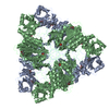
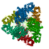
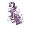
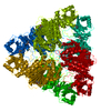
| 登録情報 | データベース: PDB / ID: 1bpm | ||||||
|---|---|---|---|---|---|---|---|
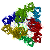
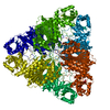
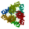
| タイトル | DIFFERENTIATION AND IDENTIFICATION OF THE TWO CATALYTIC METAL BINDING SITES IN BOVINE LENS LEUCINE AMINOPEPTIDASE BY X-RAY CRYSTALLOGRAPHY | ||||||
 要素 要素 | LEUCINE AMINOPEPTIDASE | ||||||
 キーワード キーワード | HYDROLASE(ALPHA-AMINOACYLPEPTIDE) | ||||||
| 機能・相同性 |  機能・相同性情報 機能・相同性情報cysteinylglycine-S-conjugate dipeptidase / prolyl aminopeptidase / leucyl aminopeptidase / dipeptidase activity / metalloaminopeptidase activity / carboxypeptidase activity / disordered domain specific binding / peptidase activity / manganese ion binding / mitochondrion ...cysteinylglycine-S-conjugate dipeptidase / prolyl aminopeptidase / leucyl aminopeptidase / dipeptidase activity / metalloaminopeptidase activity / carboxypeptidase activity / disordered domain specific binding / peptidase activity / manganese ion binding / mitochondrion / proteolysis / cytoplasm 類似検索 - 分子機能 | ||||||
| 生物種 |  | ||||||
| 手法 |  X線回折 / 解像度: 2.9 Å X線回折 / 解像度: 2.9 Å | ||||||
 データ登録者 データ登録者 | Kim, H. / Lipscomb, W.N. | ||||||
 引用 引用 | ジャーナル: Proc.Natl.Acad.Sci.USA / 年: 1993 タイトル: Differentiation and identification of the two catalytic metal binding sites in bovine lens leucine aminopeptidase by x-ray crystallography. 著者: Kim, H. / Lipscomb, W.N. #1: ジャーナル: J.Mol.Biol. / 年: 1992タイトル: Structure Determination and Refinement of Bovine Lens Leucine Aminopeptidase and its Complex with Bestatin 著者: Burley, S.K. / David, P.R. / Sweet, R.M. / Taylor, A. / Lipscomb, W.N. #2: ジャーナル: Proc.Natl.Acad.Sci.USA / 年: 1990タイトル: Molecular Structure of Leucine Aminopeptidase at 2.7-Angstroms Resolution 著者: Burley, S.K. / David, P.R. / Taylor, A. / Lipscomb, W.N. | ||||||
| 履歴 |
|
- 構造の表示
構造の表示
| 構造ビューア | 分子: MolmilJmol/JSmol |
|---|
- ダウンロードとリンク
-ダウンロード
| PDBx/mmCIF形式 | 1bpm.cif.gz | 101.1 KB | 表示 | PDBx/mmCIF形式 |
|---|---|---|---|---|
| PDB形式 | pdb1bpm.ent.gz | 78.4 KB | 表示 | PDB形式 |
| PDBx/mmJSON形式 | 1bpm.json.gz | ツリー表示 | PDBx/mmJSON形式 | |
| その他 |  その他のダウンロード その他のダウンロード |
-検証レポート
| アーカイブディレクトリ | https://data.pdbj.org/pub/pdb/validation_reports/bp/1bpmftp://data.pdbj.org/pub/pdb/validation_reports/bp/1bpm | HTTPS FTP |
|---|
-関連構造データ


-リンク
 PDBj
PDBj- 集合体
集合体
| 登録構造単位 | 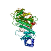
| ||||||||
|---|---|---|---|---|---|---|---|---|---|
| 1 | x 6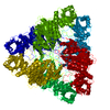
| ||||||||
| 単位格子 |
| ||||||||
| Atom site foot note | 1: RESIDUE 471 IS A CIS PROLINE. |
-要素
| #1: タンパク質 | 分子量: 52942.098 Da / 分子数: 1 / 由来タイプ: 組換発現 / 由来: (組換発現) |
|---|---|
| #2: 化合物 | ChemComp-MG /   分子量: 24.305 Da / 分子数: 1 / 由来タイプ: 合成 / 式: Mg 分子量: 24.305 Da / 分子数: 1 / 由来タイプ: 合成 / 式: Mg |
| #3: 化合物 | ChemComp-ZN /   分子量: 65.409 Da / 分子数: 1 / 由来タイプ: 合成 / 式: Zn 分子量: 65.409 Da / 分子数: 1 / 由来タイプ: 合成 / 式: Zn |
-実験情報
-実験
| 実験 | 手法: X線回折 |
|---|
- 試料調製
試料調製
| 結晶 | マシュー密度: 2.73 Å3/Da / 溶媒含有率: 54.92 % | ||||||||||||||||||||||||||||||
|---|---|---|---|---|---|---|---|---|---|---|---|---|---|---|---|---|---|---|---|---|---|---|---|---|---|---|---|---|---|---|---|
| 結晶化 | *PLUS 手法: 蒸気拡散法, ハンギングドロップ法 | ||||||||||||||||||||||||||||||
| 溶液の組成 | *PLUS
|
-データ収集
| 反射 | *PLUS 最高解像度: 2.9 Å / 最低解像度: 30 Å / Num. obs: 13523 / % possible obs: 98 % / Rmerge(I) obs: 0.162 / Num. measured all: 134829 |
|---|
- 解析
解析
| ソフトウェア | 名称: X-PLOR / 分類: 精密化 | ||||||||||||
|---|---|---|---|---|---|---|---|---|---|---|---|---|---|
| 精密化 | Rfactor Rwork: 0.189 / 最高解像度: 2.9 Å 詳細: ONE OF THE ACTIVE SITE ZINC IONS HAS BEEN REPLACED BY A MAGNESIUM ION AS DETERMINED FROM X-RAY DIFFRACTION DATA COLLECTED AT -150 DEGREES C. | ||||||||||||
| 精密化ステップ | サイクル: LAST / 最高解像度: 2.9 Å
| ||||||||||||
| 拘束条件 |
| ||||||||||||
| 精密化 | *PLUS 最高解像度: 2.9 Å / 最低解像度: 10 Å / Num. reflection obs: 10797 / σ(I): 2 / Rfactor obs: 0.189 | ||||||||||||
| 溶媒の処理 | *PLUS | ||||||||||||
| 原子変位パラメータ | *PLUS | ||||||||||||
| 拘束条件 | *PLUS
|
