 Movie
Movie Controller

[English] 日本語
 Yorodumi
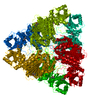
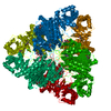
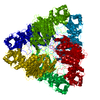
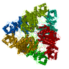
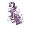
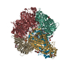
- PDB-1lcp: BOVINE LENS LEUCINE AMINOPEPTIDASE COMPLEXED WITH L-LEUCINE PHOSP... -
+ Open data

- Basic information
| Entry | Database: PDB / ID: 1lcp | ||||||
|---|---|---|---|---|---|---|---|
| Title | BOVINE LENS LEUCINE AMINOPEPTIDASE COMPLEXED WITH L-LEUCINE PHOSPHONIC ACID | ||||||
 Components Components | LEUCINE AMINOPEPTIDASE | ||||||
 Keywords Keywords | HYDROLASE (ALPHA-AMINOACYLPEPTIDE) | ||||||
| Function / homology |  Function and homology information Function and homology informationcysteinylglycine-S-conjugate dipeptidase / prolyl aminopeptidase / leucyl aminopeptidase / dipeptidase activity / metalloaminopeptidase activity / carboxypeptidase activity / disordered domain specific binding / peptidase activity / manganese ion binding / mitochondrion ...cysteinylglycine-S-conjugate dipeptidase / prolyl aminopeptidase / leucyl aminopeptidase / dipeptidase activity / metalloaminopeptidase activity / carboxypeptidase activity / disordered domain specific binding / peptidase activity / manganese ion binding / mitochondrion / proteolysis / cytoplasm Similarity search - Function | ||||||
| Biological species |  | ||||||
| Method |  X-RAY DIFFRACTION / Resolution: 1.65 Å X-RAY DIFFRACTION / Resolution: 1.65 Å | ||||||
 Authors Authors | Straeter, N. / Lipscomb, W.N. | ||||||
 Citation Citation | Journal: Biochemistry / Year: 1995 Title: Transition state analogue L-leucinephosphonic acid bound to bovine lens leucine aminopeptidase: X-ray structure at 1.65 A resolution in a new crystal form. Authors: Strater, N. / Lipscomb, W.N. #1: Journal: Adv.Enzymol.Relat.Areas Mol.Biol. / Year: 1994Title: Structure and Mechanism of Bovine Lens Leucine Aminopeptidase Authors: Kim, H. / Lipscomb, W.N. #2: Journal: Biochemistry / Year: 1993Title: X-Ray Crystallographic Determination of the Structure of Bovine Lens Leucine Aminopeptidase Complexed with Amastatin: Formulation of a Catalytic Mechanism Featuring a Gem-Diolate Transition State Authors: Kim, H. / Lipscomb, W.N. #3: Journal: Proc.Natl.Acad.Sci.USA / Year: 1993Title: Differentiation and Identification of the Two Catalytic Metal Binding Sites in Bovine Lens Leucine Aminopeptidase by X-Ray Crystallography Authors: Kim, H. / Lipscomb, W.N. #4: Journal: J.Mol.Biol. / Year: 1992Title: Structure Determination and Refinement of Bovine Lens Leucine Aminopeptidase and its Complex with Bestatin Authors: Burley, S.K. / David, P.R. / Sweet, R.M. / Taylor, A. / Lipscomb, W.N. #5: Journal: Proc.Natl.Acad.Sci.USA / Year: 1991Title: Leucine Aminopeptidase: Bestatin Inhibition and a Model for Enzyme-Catalyzed Peptide Hydrolysis Authors: Burley, S.K. / David, P.R. / Lipscomb, W.N. #6: Journal: Proc.Natl.Acad.Sci.USA / Year: 1990Title: Molecular Structure of Leucine Aminopeptidase at 2.7 Angstroms Resolution Authors: Burley, S.K. / David, P.R. / Taylor, A. / Lipscomb, W.N. | ||||||
| History |
|
- Structure visualization
| Structure viewer | Molecule: MolmilJmol/JSmol |
|---|
- Downloads & links
-Download
| PDBx/mmCIF format | 1lcp.cif.gz | 217.7 KB | Display | PDBx/mmCIF format |
|---|---|---|---|---|
| PDB format | pdb1lcp.ent.gz | 173.2 KB | Display | PDB format |
| PDBx/mmJSON format | 1lcp.json.gz | Tree view | PDBx/mmJSON format | |
| Others |  Other downloads Other downloads |
-Validation report
| Arichive directory | https://data.pdbj.org/pub/pdb/validation_reports/lc/1lcpftp://data.pdbj.org/pub/pdb/validation_reports/lc/1lcp | HTTPS FTP |
|---|
-Related structure data
| Similar structure data |
|---|

-Links
 PDBj
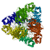
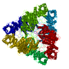
PDBj- Assembly
| Deposited unit | 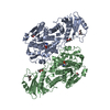
| |||||||||
|---|---|---|---|---|---|---|---|---|---|---|
| 1 | 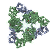
| |||||||||
| Unit cell |
| |||||||||
| Atom site foot note | 1: CIS PROLINE - PRO A 471 / 2: CIS PROLINE - PRO B 471 | |||||||||
| Components on special symmetry positions |
| |||||||||
| Noncrystallographic symmetry (NCS) | NCS oper: (Code: given Matrix: (-0.998453, -0.054621, 0.010423), Vector: Details | MTRIX THE TRANSFORMATIONS PRESENTED ON MTRIX RECORDS BELOW DESCRIBE NON-CRYSTALLOGRAPHIC RELATIONSHIPS AMONG THE VARIOUS DOMAINS IN THIS ENTRY. APPLYING THE APPROPRIATE MTRIX TRANSFORMATION TO THE RESIDUES LISTED FIRST WILL YIELD APPROXIMATE COORDINATES FOR THE RESIDUES LISTED SECOND. APPLIED TO TRANSFORMED TO MTRIX RESIDUES RESIDUES RMSD M1 B 1 .. 484 A 1 .. 484 0.218 SYMMETRY THE CRYSTALLOGRAPHIC SYMMETRY TRANSFORMATIONS PRESENTED BELOW GENERATE THE SUBUNITS OF THE POLYMERIC MOLECULE. APPLIED TO RESIDUES: A 1 .. 490 1 OF 4 TRANSFORMATIONS TO GENERATE ONE HEXAMER OF 32 SYMMETRY. SYMMETRY1 1 -0.500000 -0.866025 0.000000 65.15000 SYMMETRY2 1 0.866025 -0.500000 0.000000 112.84310 SYMMETRY3 1 0.000000 0.000000 1.000000 0.00000 APPLIED TO RESIDUES: A 1 .. 490 1 OF 4 TRANSFORMATIONS TO GENERATE ONE HEXAMER OF 32 SYMMETRY. SYMMETRY1 2 -0.500000 0.866025 0.000000 -65.15000 SYMMETRY2 2 -0.866025 -0.500000 0.000000 112.84310 SYMMETRY3 2 0.000000 0.000000 1.000000 0.00000 APPLIED TO RESIDUES: B 1 .. 490 1 OF 4 TRANSFORMATIONS TO GENERATE ONE HEXAMER OF 32 SYMMETRY. SYMMETRY1 1 -0.500000 -0.866025 0.000000 65.15000 SYMMETRY2 1 0.866025 -0.500000 0.000000 112.84310 SYMMETRY3 1 0.000000 0.000000 1.000000 0.00000 APPLIED TO RESIDUES: B 1 .. 490 1 OF 4 TRANSFORMATIONS TO GENERATE ONE HEXAMER OF 32 SYMMETRY. SYMMETRY1 2 -0.500000 0.866025 0.000000 -65.15000 SYMMETRY2 2 -0.866025 -0.500000 0.000000 112.84310 SYMMETRY3 2 0.000000 0.000000 1.000000 0.00000 | |
-Components
| #1: Protein | Mass: 52668.855 Da / Num. of mol.: 2 / Source method: isolated from a natural source / Source: (natural) #2: Chemical | ChemComp-ZN /   Mass: 65.409 Da / Num. of mol.: 6 / Source method: obtained synthetically / Formula: Zn Mass: 65.409 Da / Num. of mol.: 6 / Source method: obtained synthetically / Formula: Zn#3: Chemical |   Mass: 167.143 Da / Num. of mol.: 2 / Source method: obtained synthetically / Formula: C5H14NO3P Mass: 167.143 Da / Num. of mol.: 2 / Source method: obtained synthetically / Formula: C5H14NO3P#4: Chemical | ChemComp-MRD / (   Mass: 118.174 Da / Num. of mol.: 6 / Source method: obtained synthetically / Formula: C6H14O2 / Comment: precipitant*YM Mass: 118.174 Da / Num. of mol.: 6 / Source method: obtained synthetically / Formula: C6H14O2 / Comment: precipitant*YM#5: Water | ChemComp-HOH / |  Mass: 18.015 Da / Num. of mol.: 968 / Source method: isolated from a natural source / Formula: H2O Mass: 18.015 Da / Num. of mol.: 968 / Source method: isolated from a natural source / Formula: H2OCompound details | THE SECONDARY STRUCTURE ASSIGNMENT IS TAKEN FROM REFERENCE 3. THERE ARE NO SIGNIFICANT DEVIATIONS ...THE SECONDARY STRUCTURE ASSIGNMENT | |
|---|
-Experimental details
-Experiment
| Experiment | Method: X-RAY DIFFRACTION / Number of used crystals: 1 |
|---|
- Sample preparation
| Crystal | Density Matthews: 2.92 Å3/Da / Density % sol: 57.88 % | ||||||||||||||||||||||||||||||||||||||||||||||||||||||
|---|---|---|---|---|---|---|---|---|---|---|---|---|---|---|---|---|---|---|---|---|---|---|---|---|---|---|---|---|---|---|---|---|---|---|---|---|---|---|---|---|---|---|---|---|---|---|---|---|---|---|---|---|---|---|---|
| Crystal grow | *PLUS pH: 7.8 / Method: vapor diffusion, hanging drop | ||||||||||||||||||||||||||||||||||||||||||||||||||||||
| Components of the solutions | *PLUS
|
-Data collection
| Diffraction source | Wavelength: 1.5418 Å |
|---|---|
| Detector | Type: XENTRONICS / Detector: AREA DETECTOR / Date: Dec 8, 1994 |
| Radiation | Monochromator: SUPPER DOUBLE MIRROR / Monochromatic (M) / Laue (L): M / Scattering type: x-ray |
| Radiation wavelength | Wavelength: 1.5418 Å / Relative weight: 1 |
| Reflection | Resolution: 1.65→36 Å / Num. obs: 144211 / % possible obs: 97.7 % / Observed criterion σ(I): 0 / Redundancy: 5.3 % / Rmerge(I) obs: 0.074 |
| Reflection | *PLUS Num. measured all: 766198 / Rmerge(I) obs: 0.074 |
- Processing
| Software |
| ||||||||||||||||
|---|---|---|---|---|---|---|---|---|---|---|---|---|---|---|---|---|---|
| Refinement | Resolution: 1.65→7 Å / σ(F): 2 Details: ALL WATER MOLECULES EXCEPT OF 4 HAVE BEEN REFINED WITH THE PARAM19.SOL PARAMETER FILE IN X-PLOR 3.1. THE FOUR ACTIVE SITE WATER MOLECULES 967, 968, 969, AND 970 HAVE BEEN REFINED TURNING OFF ...Details: ALL WATER MOLECULES EXCEPT OF 4 HAVE BEEN REFINED WITH THE PARAM19.SOL PARAMETER FILE IN X-PLOR 3.1. THE FOUR ACTIVE SITE WATER MOLECULES 967, 968, 969, AND 970 HAVE BEEN REFINED TURNING OFF NON-BONDED INTERACTIONS WITH EACH OTHER IN ORDER TO GET A NON-BIASED VALUE OF THE SHORT DISTANCE BETWEEN WATER MOLECULES 967 AND 968 (2.3 ANG) AND BETWEEN 969 AND 970 (2.1 ANG).
| ||||||||||||||||
| Displacement parameters | Biso mean: 12.7 Å2 | ||||||||||||||||
| Refine analyze | Luzzati coordinate error obs: 0.17 Å | ||||||||||||||||
| Refinement step | Cycle: LAST / Resolution: 1.65→7 Å
| ||||||||||||||||
| Software | *PLUS Name: X-PLOR / Classification: refinement | ||||||||||||||||
| Refinement | *PLUS Rfactor obs: 0.16 / Rfactor Rwork: 0.16 | ||||||||||||||||
| Solvent computation | *PLUS | ||||||||||||||||
| Displacement parameters | *PLUS | ||||||||||||||||
| Refine LS restraints | *PLUS
|
