[English] 日本語
Yorodumi
- PDB-1ao5: MOUSE GLANDULAR KALLIKREIN-13 (PRORENIN CONVERTING ENZYME) -

+
Open data


ID or keywords:

Loading...

-
Basic information

Entry
Database: PDB / ID: 1ao5
TitleMOUSE GLANDULAR KALLIKREIN-13 (PRORENIN CONVERTING ENZYME)
ComponentsGLANDULAR KALLIKREIN-13
Keywords
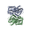
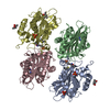
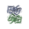
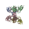
SERINE PROTEASE / GLANDULAR KALLIKREIN / PROTEIN MATURATION
Function / homology
Function and homology information


kallikrein 13 / regulation of systemic arterial blood pressure / zymogen activation / secretory granule / peptidase activity / serine-type endopeptidase activity / extracellular space
Similarity search - Function
Serine proteases, trypsin family, histidine active site / Peptidase S1A, chymotrypsin family / Serine proteases, trypsin family, histidine active site. / Serine proteases, trypsin domain profile. / Trypsin-like serine protease / Serine proteases, trypsin domain / Trypsin / Trypsin-like serine proteases / Thrombin, subunit H / Peptidase S1, PA clan, chymotrypsin-like fold ...Serine proteases, trypsin family, histidine active site / Peptidase S1A, chymotrypsin family / Serine proteases, trypsin family, histidine active site. / Serine proteases, trypsin domain profile. / Trypsin-like serine protease / Serine proteases, trypsin domain / Trypsin / Trypsin-like serine proteases / Thrombin, subunit H / Peptidase S1, PA clan, chymotrypsin-like fold / Peptidase S1, PA clan / Beta Barrel / Mainly Beta
Similarity search - Domain/homology
Epidermal growth factor-binding protein type B
Similarity search - Component
Biological speciesMus musculus (house mouse)
MethodX-RAY DIFFRACTION / SYNCHROTRON / MOLECULAR REPLACEMENT / Resolution: 2.6 Å
AuthorsTimm, D.E.
CitationJournal: Protein Sci. / Year: 1997
Title: The crystal structure of the mouse glandular kallikrein-13 (prorenin converting enzyme)
Authors: Timm, D.E.
History
DepositionJul 16, 1997Processing site: BNL
Revision 1.0Oct 15, 1997Provider: repository / Type: Initial release
Revision 1.1Mar 3, 2008Group: Version format compliance
Revision 1.2Jul 13, 2011Group: Non-polymer description / Version format compliance
Revision 2.0Jul 29, 2020Group: Atomic model / Data collection ...Atomic model / Data collection / Database references / Derived calculations / Structure summary
Category: atom_site / chem_comp ...atom_site / chem_comp / entity / pdbx_branch_scheme / pdbx_chem_comp_identifier / pdbx_entity_branch / pdbx_entity_branch_descriptor / pdbx_entity_branch_link / pdbx_entity_branch_list / pdbx_entity_nonpoly / pdbx_nonpoly_scheme / pdbx_struct_assembly_gen / struct_asym / struct_conn / struct_ref_seq_dif / struct_site / struct_site_gen
Item: _atom_site.auth_asym_id / _atom_site.auth_seq_id ..._atom_site.auth_asym_id / _atom_site.auth_seq_id / _atom_site.label_asym_id / _chem_comp.name / _chem_comp.type / _entity.formula_weight / _entity.pdbx_description / _entity.pdbx_number_of_molecules / _entity.type / _pdbx_struct_assembly_gen.asym_id_list / _struct_conn.pdbx_dist_value / _struct_conn.pdbx_leaving_atom_flag / _struct_conn.pdbx_role / _struct_conn.ptnr1_auth_asym_id / _struct_conn.ptnr1_auth_comp_id / _struct_conn.ptnr1_auth_seq_id / _struct_conn.ptnr1_label_asym_id / _struct_conn.ptnr1_label_atom_id / _struct_conn.ptnr1_label_comp_id / _struct_conn.ptnr1_label_seq_id / _struct_conn.ptnr2_auth_asym_id / _struct_conn.ptnr2_auth_comp_id / _struct_conn.ptnr2_auth_seq_id / _struct_conn.ptnr2_label_asym_id / _struct_conn.ptnr2_label_atom_id / _struct_conn.ptnr2_label_comp_id / _struct_conn.ptnr2_label_seq_id / _struct_ref_seq_dif.details
Description: Carbohydrate remediation / Provider: repository / Type: Remediation
Revision 2.1Aug 2, 2023Group: Database references / Refinement description / Structure summary
Category: chem_comp / database_2 / pdbx_initial_refinement_model
Item: _chem_comp.pdbx_synonyms / _database_2.pdbx_DOI / _database_2.pdbx_database_accession

-
Structure visualization

Structure viewerMolecule:
MolmilJmol/JSmol

Downloads & links

-
Assembly

Deposited unit
A: GLANDULAR KALLIKREIN-13
B: GLANDULAR KALLIKREIN-13
hetero molecules


Theoretical massNumber of molelcules
Total (without water)53,0614
Polymers52,2122
Non-polymers8492
Water2,144119
1
A: GLANDULAR KALLIKREIN-13
B: GLANDULAR KALLIKREIN-13
hetero molecules

A: GLANDULAR KALLIKREIN-13
B: GLANDULAR KALLIKREIN-13
hetero molecules


Theoretical massNumber of molelcules
Total (without water)106,1228
Polymers104,4254
Non-polymers1,6984
Water724
TypeNameSymmetry operationNumber
identity operation1_555x,y,z1
crystal symmetry operation7_556y,x,-z+11
Unit cell
Length a, b, c (Å)73.750, 73.750, 192.710
Angle α, β, γ (deg.)90.00, 90.00, 90.00
Int Tables number96
Space group name H-MP43212
Noncrystallographic symmetry (NCS)NCS oper:
IDCodeMatrixVector
1given(-0.967743, -0.233481, -0.094661), (-0.249462, 0.940587, 0.230358), (0.035252, 0.246542, -0.968491)45.18026, -13.42768, 167.78738
2given(-0.971978, -0.23159, -0.040316), (-0.234519, 0.943552, 0.233902), (-0.016129, 0.236802, -0.971424)40.89381, -13.77748, 168.20828

-
Components

#1: Protein GLANDULAR KALLIKREIN-13 / PRORENIN CONVERTING ENZYME (PRECE) / EPIDERMAL GROWTH FACTOR BINDING PROTEIN TYPE B (EGF-BP B)


Mass: 26106.150 Da / Num. of mol.: 2 / Source method: isolated from a natural source / Source: (natural) Mus musculus (house mouse) / Organ: SUBMANDIBULAR GLAND / Strain: SWISS WEBSTER / References: UniProt: P36368, tissue kallikrein
#2: Polysaccharide 2-acetamido-2-deoxy-beta-D-glucopyranose-(1-4)-2-acetamido-2-deoxy-beta-D-glucopyranose


Type: oligosaccharide / Mass: 424.401 Da / Num. of mol.: 2
Source method: isolated from a genetically manipulated source
DescriptorTypeProgram
DGlcpNAcb1-4DGlcpNAcb1-Glycam Condensed SequenceGMML 1.0
WURCS=2.0/1,2,1/[a2122h-1b_1-5_2*NCC/3=O]/1-1/a4-b1WURCSPDB2Glycan 1.1.0
[]{[(4+1)][b-D-GlcpNAc]{[(4+1)][b-D-GlcpNAc]{}}}LINUCSPDB-CARE
#3: Water ChemComp-HOH / water / Water


Mass: 18.015 Da / Num. of mol.: 119 / Source method: isolated from a natural source / Formula: H2O
Sequence detailsTHE NUMBERING OF RESIDUES IN PDB ENTRY IS RELATIVE TO CHYMOTRYPSIN.

-
Experimental details

-
Experiment

ExperimentMethod: X-RAY DIFFRACTION / Number of used crystals: 1

-
Sample preparation

CrystalDensity Matthews: 2.51 Å3/Da / Density % sol: 47.7 %
Crystal growMethod: vapor diffusion, hanging drop / pH: 6.5
Details: MGK-13 (57 MG/ML) WAS CRYSTALLIZED USING 10-15% PEG8000, 0.2M LISO4, 0.1M SODIUM CACODYLATE, PH 6.5 BY HANGING DROP VAPOR DIFFUSION METHOD., vapor diffusion - hanging drop
Crystal grow
*PLUS
Method: vapor diffusion, hanging drop
Components of the solutions
*PLUS
IDConc.Common nameCrystal-IDSol-IDChemical formula
157 mg/mlmGK-131drop
210-15 %PEG80001reservoir
3200 mM1reservoirLi2SO4
4100 mMsodium cacodylate1reservoir

-
Data collection

DiffractionMean temperature: 277 K
Diffraction sourceSource: SYNCHROTRON / Site: SRS / Beamline: PX9.6 / Wavelength: 0.87
DetectorType: MARRESEARCH / Detector: IMAGE PLATE / Date: Jul 1, 1995 / Details: SYNCHROTRON
RadiationMonochromator: SYNCHROTRON / Monochromatic (M) / Laue (L): M / Scattering type: x-ray
Radiation wavelengthWavelength: 0.87 Å / Relative weight: 1
ReflectionResolution: 2.55→40.33 Å / Num. obs: 17034 / % possible obs: 95.3 % / Observed criterion σ(I): 2 / Redundancy: 2.5 % / Rmerge(I) obs: 0.065 / Rsym value: 0.065 / Net I/σ(I): 9.6
Reflection shellResolution: 2.55→2.69 Å / Redundancy: 2.3 % / Rmerge(I) obs: 0.345 / Mean I/σ(I) obs: 1.7 / Rsym value: 0.345 / % possible all: 86.8

-
Processing

Software
NameVersionClassification
MOSFLMdata reduction
CCP4data reduction
X-PLOR3.1model building
X-PLOR3.1refinement
CCP4data scaling
X-PLOR3.1phasing
RefinementMethod to determine structure: MOLECULAR REPLACEMENT
Starting model: PDB ENTRY 2KAI
Resolution: 2.6→40.33 Å / Data cutoff high absF: 100000 / Data cutoff low absF: 0.1 / Isotropic thermal model: GROUPED / Cross valid method: THROUGHOUT / σ(F): 2
Details: A BULK SOLVENT CORRECTION WAS APPLIED USING X-PLOR V3.1. RESIDUES 16 - 246 (CHAINS A AND B) REPRESENT THE TWO MOLECULES PRESENT IN THE ASYMMETRIC UNIT. A CHAIN BREAK OCCURS IN EACH MOLECULE ...Details: A BULK SOLVENT CORRECTION WAS APPLIED USING X-PLOR V3.1. RESIDUES 16 - 246 (CHAINS A AND B) REPRESENT THE TWO MOLECULES PRESENT IN THE ASYMMETRIC UNIT. A CHAIN BREAK OCCURS IN EACH MOLECULE AT THR 147. RESIDUE ARG 148 AT A PROTEOLYTIC CLEAVAGE SITE IS DISORDERED AND MISSING FROM THE ELECTRON DENSITY.
RfactorNum. reflection% reflectionSelection details
Rfree0.271 785 5 %RANDOM
Rwork0.197 ---
obs0.197 16364 95.3 %-
Refinement stepCycle: LAST / Resolution: 2.6→40.33 Å
ProteinNucleic acidLigandSolventTotal
Num. atoms3630 0 56 119 3805
Refine LS restraints
Refine-IDTypeDev ideal
X-RAY DIFFRACTIONx_bond_d0.008
X-RAY DIFFRACTIONx_bond_d_na
X-RAY DIFFRACTIONx_bond_d_prot
X-RAY DIFFRACTIONx_angle_d
X-RAY DIFFRACTIONx_angle_d_na
X-RAY DIFFRACTIONx_angle_d_prot
X-RAY DIFFRACTIONx_angle_deg1.74
X-RAY DIFFRACTIONx_angle_deg_na
X-RAY DIFFRACTIONx_angle_deg_prot
X-RAY DIFFRACTIONx_dihedral_angle_d24.8
X-RAY DIFFRACTIONx_dihedral_angle_d_na
X-RAY DIFFRACTIONx_dihedral_angle_d_prot
X-RAY DIFFRACTIONx_improper_angle_d1.279
X-RAY DIFFRACTIONx_improper_angle_d_na
X-RAY DIFFRACTIONx_improper_angle_d_prot
X-RAY DIFFRACTIONx_mcbond_it
X-RAY DIFFRACTIONx_mcangle_it
X-RAY DIFFRACTIONx_scbond_it
X-RAY DIFFRACTIONx_scangle_it
Refine LS restraints NCSNCS model details: RESTRAINTS
LS refinement shellResolution: 2.6→2.72 Å / Total num. of bins used: 8
RfactorNum. reflection% reflection
Rfree0.3606 55 2.8 %
Rwork0.3317 1895 -
Xplor file
Refine-IDSerial noParam file
X-RAY DIFFRACTION1PARHCSDX.PRO
X-RAY DIFFRACTION2PARAM19.SOL
X-RAY DIFFRACTION3PARAM3_MOD.CHO
Software
*PLUS
Name: X-PLOR / Version: 3.1 / Classification: refinement
Refine LS restraints
*PLUS
Refine-IDTypeDev ideal
X-RAY DIFFRACTIONx_dihedral_angle_d
X-RAY DIFFRACTIONx_dihedral_angle_deg24.808
X-RAY DIFFRACTIONx_improper_angle_d
X-RAY DIFFRACTIONx_improper_angle_deg1.279

+
About Yorodumi

-
News

-
Feb 9, 2022. New format data for meta-information of EMDB entries

New format data for meta-information of EMDB entries

  • Version 3 of the EMDB header file is now the official format.
  • The previous official version 1.9 will be removed from the archive.

Related info.:EMDB header

External links:wwPDB to switch to version 3 of the EMDB data model

-
Aug 12, 2020. Covid-19 info

Covid-19 info

URL: https://pdbj.org/emnavi/covid19.php

New page: Covid-19 featured information page in EM Navigator.

Related info.:Covid-19 info / Mar 5, 2020. Novel coronavirus structure data

+
Mar 5, 2020. Novel coronavirus structure data

Novel coronavirus structure data

Related info.:Yorodumi Speices / Aug 12, 2020. Covid-19 info

External links:COVID-19 featured content - PDBj / Molecule of the Month (242):Coronavirus Proteases

+
Jan 31, 2019. EMDB accession codes are about to change! (news from PDBe EMDB page)

EMDB accession codes are about to change! (news from PDBe EMDB page)

  • The allocation of 4 digits for EMDB accession codes will soon come to an end. Whilst these codes will remain in use, new EMDB accession codes will include an additional digit and will expand incrementally as the available range of codes is exhausted. The current 4-digit format prefixed with “EMD-” (i.e. EMD-XXXX) will advance to a 5-digit format (i.e. EMD-XXXXX), and so on. It is currently estimated that the 4-digit codes will be depleted around Spring 2019, at which point the 5-digit format will come into force.
  • The EM Navigator/Yorodumi systems omit the EMD- prefix.

Related info.:Q: What is EMD? / ID/Accession-code notation in Yorodumi/EM Navigator

External links:EMDB Accession Codes are Changing Soon! / Contact to PDBj

+
Jul 12, 2017. Major update of PDB

Major update of PDB

  • wwPDB released updated PDB data conforming to the new PDBx/mmCIF dictionary.
  • This is a major update changing the version number from 4 to 5, and with Remediation, in which all the entries are updated.
  • In this update, many items about electron microscopy experimental information are reorganized (e.g. em_software).
  • Now, EM Navigator and Yorodumi are based on the updated data.

External links:wwPDB Remediation / Enriched Model Files Conforming to OneDep Data Standards Now Available in the PDB FTP Archive

-
Yorodumi

Thousand views of thousand structures

  • Yorodumi is a browser for structure data from EMDB, PDB, SASBDB, etc.
  • This page is also the successor to EM Navigator detail page, and also detail information page/front-end page for Omokage search.
  • The word "yorodu" (or yorozu) is an old Japanese word meaning "ten thousand". "mi" (miru) is to see.

Related info.:EMDB / PDB / SASBDB / Comparison of 3 databanks / Yorodumi Search / Aug 31, 2016. New EM Navigator & Yorodumi / Yorodumi Papers / Jmol/JSmol / Function and homology information / Changes in new EM Navigator and Yorodumi

Read more