 Movie
Movie Controller


+ Open data

- Basic information
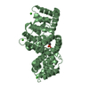
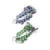
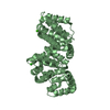
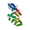
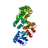
| Entry | Database: PDB / ID: 1ain | ||||||
|---|---|---|---|---|---|---|---|
| Title | CRYSTAL STRUCTURE OF HUMAN ANNEXIN I AT 2.5 ANGSTROMS RESOLUTION | ||||||
 Components Components | ANNEXIN I | ||||||
 Keywords Keywords | CALCIUM/PHOSPHOLIPID BINDING / CALCIUM-PHOSPHOLIPID BINDING complex | ||||||
| Function / homology |  Function and homology information Function and homology informationregulation of interleukin-1 production / prolactin secretion / myoblast migration involved in skeletal muscle regeneration / regulation of leukocyte migration / granulocyte chemotaxis / regulation of hormone secretion / positive regulation of T-helper 1 cell differentiation / phospholipase A2 inhibitor activity / positive regulation of vesicle fusion / neutrophil clearance ...regulation of interleukin-1 production / prolactin secretion / myoblast migration involved in skeletal muscle regeneration / regulation of leukocyte migration / granulocyte chemotaxis / regulation of hormone secretion / positive regulation of T-helper 1 cell differentiation / phospholipase A2 inhibitor activity / positive regulation of vesicle fusion / neutrophil clearance / prostate gland development / negative regulation of T-helper 2 cell differentiation / positive regulation of prostaglandin biosynthetic process / endocrine pancreas development / negative regulation of interleukin-8 production / neutrophil activation / cadherin binding involved in cell-cell adhesion / peptide cross-linking / cornified envelope / positive regulation of neutrophil apoptotic process / gliogenesis / neutrophil homeostasis / hepatocyte differentiation / calcium-dependent phospholipid binding / Formyl peptide receptors bind formyl peptides and many other ligands / negative regulation of exocytosis / motile cilium / cellular response to glucocorticoid stimulus / insulin secretion / alpha-beta T cell differentiation / DNA duplex unwinding / phagocytic cup / arachidonic acid secretion / positive regulation of cell migration involved in sprouting angiogenesis / positive regulation of wound healing / G protein-coupled receptor signaling pathway, coupled to cyclic nucleotide second messenger / monocyte chemotaxis / response to X-ray / lateral plasma membrane / Smooth Muscle Contraction / positive regulation of G1/S transition of mitotic cell cycle / cellular response to vascular endothelial growth factor stimulus / localization / phagocytosis / keratinocyte differentiation / positive regulation of T cell proliferation / positive regulation of interleukin-2 production / response to interleukin-1 / phospholipid binding / adherens junction / sarcolemma / response to peptide hormone / cellular response to hydrogen peroxide / calcium-dependent protein binding / response to estradiol / regulation of cell shape / G alpha (i) signalling events / regulation of inflammatory response / actin cytoskeleton organization / early endosome membrane / G alpha (q) signalling events / basolateral plasma membrane / collagen-containing extracellular matrix / Interleukin-4 and Interleukin-13 signaling / vesicle / adaptive immune response / cell surface receptor signaling pathway / endosome / inflammatory response / apical plasma membrane / signaling receptor binding / focal adhesion / innate immune response / lipid binding / calcium ion binding / negative regulation of apoptotic process / cell surface / signal transduction / extracellular space / extracellular exosome / extracellular region / nucleoplasm / nucleus / plasma membrane / cytosol / cytoplasmSimilarity search - Function | ||||||
| Biological species |  Homo sapiens (human) Homo sapiens (human) | ||||||
| Method | X-RAY DIFFRACTION / Resolution: 2.5 Å | ||||||
 Authors Authors | Kim, S.-H. | ||||||
 Citation Citation | Journal: Protein Sci. / Year: 1993 Title: Crystal structure of human annexin I at 2.5 A resolution. Authors: Weng, X. / Luecke, H. / Song, I.S. / Kang, D.S. / Kim, S.H. / Huber, R. | ||||||
| History |
|
- Structure visualization
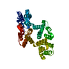
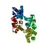
| Structure viewer | Molecule: MolmilJmol/JSmol |
|---|
- Downloads & links
-Download
| PDBx/mmCIF format | 1ain.cif.gz | 22.1 KB | Display | PDBx/mmCIF format |
|---|---|---|---|---|
| PDB format | pdb1ain.ent.gz | 10.6 KB | Display | PDB format |
| PDBx/mmJSON format | 1ain.json.gz | Tree view | PDBx/mmJSON format | |
| Others |  Other downloads Other downloads |
-Validation report
| Arichive directory | https://data.pdbj.org/pub/pdb/validation_reports/ai/1ainftp://data.pdbj.org/pub/pdb/validation_reports/ai/1ain | HTTPS FTP |
|---|
-Related structure data
| Similar structure data |
|---|


-Links
 PDBj
PDBj


- Assembly
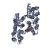
| Deposited unit | 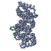
| ||||||||
|---|---|---|---|---|---|---|---|---|---|
| 1 |
| ||||||||
| Unit cell |
|
-Components
| #1: Protein | / Coordinate model: Cα atoms only Mass: 35093.121 Da / Num. of mol.: 1 Source method: isolated from a genetically manipulated source Source: (gene. exp.) Homo sapiens (human) / References: UniProt: P04083 |
|---|---|
| #2: Chemical | ChemComp-CA /   Mass: 40.078 Da / Num. of mol.: 6 / Source method: obtained synthetically / Formula: Ca Mass: 40.078 Da / Num. of mol.: 6 / Source method: obtained synthetically / Formula: Ca |
-Experimental details
-Experiment
| Experiment | Method: X-RAY DIFFRACTION |
|---|
- Sample preparation
| Crystal | Density Matthews: 2.82 Å3/Da / Density % sol: 56.4 % | ||||||||||||||||||||||||||||||||||||||||||||||||||||||||||||
|---|---|---|---|---|---|---|---|---|---|---|---|---|---|---|---|---|---|---|---|---|---|---|---|---|---|---|---|---|---|---|---|---|---|---|---|---|---|---|---|---|---|---|---|---|---|---|---|---|---|---|---|---|---|---|---|---|---|---|---|---|---|
| Crystal grow | *PLUS pH: 6.5 / Method: other | ||||||||||||||||||||||||||||||||||||||||||||||||||||||||||||
| Components of the solutions | *PLUS
|
-Data collection
| Radiation | Scattering type: x-ray |
|---|---|
| Radiation wavelength | Relative weight: 1 |
| Reflection | *PLUS Highest resolution: 2.5 Å / Num. obs: 13945 / % possible obs: 97.3 % / Rmerge(I) obs: 0.0535 / Num. measured all: 76668 |
- Processing
| Software |
| ||||||||||||
|---|---|---|---|---|---|---|---|---|---|---|---|---|---|
| Refinement | Rfactor Rwork: 0.163 / Highest resolution: 2.5 Å | ||||||||||||
| Refinement step | Cycle: LAST / Highest resolution: 2.5 Å
| ||||||||||||
| Refine LS restraints |
| ||||||||||||
| Refinement | *PLUS Highest resolution: 2.5 Å / Lowest resolution: 10 Å / Rfactor obs: 0.177 | ||||||||||||
| Solvent computation | *PLUS | ||||||||||||
| Displacement parameters | *PLUS | ||||||||||||
| Refine LS restraints | *PLUS
|
