 Movie
Movie Controller

[English] 日本語
 Yorodumi
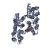
- PDB-5lq0: Crystal structure of Tyr24 phosphorylated Annexin A2 at 2.9 A res... -
+ Open data

- Basic information
| Entry | Database: PDB / ID: 5lq0 | ||||||
|---|---|---|---|---|---|---|---|
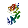
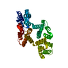
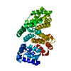
| Title | Crystal structure of Tyr24 phosphorylated Annexin A2 at 2.9 A resolution | ||||||
 Components Components | Annexin A2 | ||||||
 Keywords Keywords | calcium binding protein / tyrosine phosphorylation | ||||||
| Function / homology |  Function and homology information Function and homology informationAnxA2-p11 complex / membrane raft assembly / positive regulation of receptor-mediated endocytosis involved in cholesterol transport / positive regulation of vacuole organization / phospholipase A2 inhibitor activity / positive regulation of low-density lipoprotein particle clearance / negative regulation of low-density lipoprotein particle receptor catabolic process / positive regulation of plasma membrane repair / positive regulation of plasminogen activation / PCSK9-AnxA2 complex ...AnxA2-p11 complex / membrane raft assembly / positive regulation of receptor-mediated endocytosis involved in cholesterol transport / positive regulation of vacuole organization / phospholipase A2 inhibitor activity / positive regulation of low-density lipoprotein particle clearance / negative regulation of low-density lipoprotein particle receptor catabolic process / positive regulation of plasma membrane repair / positive regulation of plasminogen activation / PCSK9-AnxA2 complex / myelin sheath adaxonal region / cadherin binding involved in cell-cell adhesion / positive regulation of vesicle fusion / cornified envelope / Schmidt-Lanterman incisure / vesicle budding from membrane / calcium-dependent phospholipid binding / osteoclast development / negative regulation of receptor internalization / plasma membrane protein complex / Dissolution of Fibrin Clot / S100 protein binding / collagen fibril organization / epithelial cell apoptotic process / vesicle membrane / phosphatidylserine binding / positive regulation of receptor recycling / positive regulation of exocytosis / basement membrane / regulation of neurogenesis / Smooth Muscle Contraction / fibrinolysis / cytoskeletal protein binding / phosphatidylinositol-4,5-bisphosphate binding / lipid droplet / lung development / Gene and protein expression by JAK-STAT signaling after Interleukin-12 stimulation / Turbulent (oscillatory, disturbed) flow shear stress activates signaling by PIEZO1 and integrins in endothelial cells / cell-matrix adhesion / response to activity / Developmental Lineage of Pancreatic Ductal Cells / adherens junction / serine-type endopeptidase inhibitor activity / sarcolemma / mRNA transcription by RNA polymerase II / RNA polymerase II transcription regulator complex / nuclear matrix / calcium-dependent protein binding / azurophil granule lumen / melanosome / late endosome membrane / extracellular matrix / protease binding / angiogenesis / midbody / vesicle / basolateral plasma membrane / early endosome / cell adhesion / endosome / lysosomal membrane / calcium ion binding / Neutrophil degranulation / cell surface / positive regulation of transcription by RNA polymerase II / : / RNA binding / extracellular exosome / extracellular region / membrane / identical protein binding / nucleus / plasma membrane / cytoplasm / cytosol Similarity search - Function | ||||||
| Biological species |  Homo sapiens (human) Homo sapiens (human) | ||||||
| Method |  X-RAY DIFFRACTION / SYNCHROTRON / MOLECULAR REPLACEMENT / Resolution: 2.9 Å X-RAY DIFFRACTION / SYNCHROTRON / MOLECULAR REPLACEMENT / Resolution: 2.9 Å | ||||||
 Authors Authors | Ecsedi, P. / Gogl, G. / Kiss, B. / Nyitray, L. | ||||||
 Citation Citation | Journal: Structure / Year: 2017 Title: Regulation of the Equilibrium between Closed and Open Conformations of Annexin A2 by N-Terminal Phosphorylation and S100A4-Binding. Authors: Ecsedi, P. / Kiss, B. / Gogl, G. / Radnai, L. / Buday, L. / Koprivanacz, K. / Liliom, K. / Leveles, I. / Vertessy, B. / Jeszenoi, N. / Hetenyi, C. / Schlosser, G. / Katona, G. / Nyitray, L. | ||||||
| History |
|
- Structure visualization
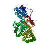
| Structure viewer | Molecule: MolmilJmol/JSmol |
|---|
- Downloads & links
-Download
| PDBx/mmCIF format | 5lq0.cif.gz | 265.4 KB | Display | PDBx/mmCIF format |
|---|---|---|---|---|
| PDB format | pdb5lq0.ent.gz | 214.7 KB | Display | PDB format |
| PDBx/mmJSON format | 5lq0.json.gz | Tree view | PDBx/mmJSON format | |
| Others |  Other downloads Other downloads |
-Validation report
| Arichive directory | https://data.pdbj.org/pub/pdb/validation_reports/lq/5lq0ftp://data.pdbj.org/pub/pdb/validation_reports/lq/5lq0 | HTTPS FTP |
|---|
-Related structure data
| Related structure data | 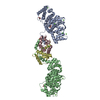 5lpuC  5lpxC  5lq2C  2hywS C: citing same article ( S: Starting model for refinement |
|---|---|
| Similar structure data |


-Links
 PDBj
PDBj


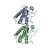
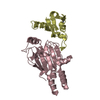
- Assembly
| Deposited unit | 
| ||||||||
|---|---|---|---|---|---|---|---|---|---|
| 1 | 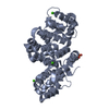
| ||||||||
| 2 | 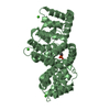
| ||||||||
| Unit cell |
|
-Components
| #1: Protein | Mass: 38693.918 Da / Num. of mol.: 2 / Mutation: A66E Y24(PTR) Source method: isolated from a genetically manipulated source Source: (gene. exp.) Homo sapiens (human) / Gene: ANXA2, ANX2, ANX2L4, CAL1H, LPC2D / Production host:  #2: Chemical | ChemComp-CA /   Mass: 40.078 Da / Num. of mol.: 6 / Source method: obtained synthetically / Formula: Ca Mass: 40.078 Da / Num. of mol.: 6 / Source method: obtained synthetically / Formula: Ca#3: Water | ChemComp-HOH / |  Mass: 18.015 Da / Num. of mol.: 7 / Source method: isolated from a natural source / Formula: H2O Mass: 18.015 Da / Num. of mol.: 7 / Source method: isolated from a natural source / Formula: H2OHas protein modification | Y | |
|---|
-Experimental details
-Experiment
| Experiment | Method: X-RAY DIFFRACTION / Number of used crystals: 1 |
|---|
- Sample preparation
| Crystal | Density Matthews: 3.11 Å3/Da / Density % sol: 60.48 % |
|---|---|
| Crystal grow | Temperature: 296 K / Method: vapor diffusion, hanging drop Details: 0.1 M MES pH 6, 0.1 M magnesium chloride, 4% PEG6000 |
-Data collection
| Diffraction | Mean temperature: 100 K |
|---|---|
| Diffraction source | Source: SYNCHROTRON / Site: SLS  / Beamline: X06DA / Wavelength: 1 Å / Beamline: X06DA / Wavelength: 1 Å |
| Detector | Type: DECTRIS PILATUS 2M-F / Detector: PIXEL / Date: Dec 6, 2015 |
| Radiation | Protocol: SINGLE WAVELENGTH / Monochromatic (M) / Laue (L): M / Scattering type: x-ray |
| Radiation wavelength | Wavelength: 1 Å / Relative weight: 1 |
| Reflection | Resolution: 2.9→47.77 Å / Num. obs: 22187 / % possible obs: 99.12 % / Redundancy: 9.14 % / CC1/2: 0.999 / Rsym value: 0.094 / Net I/σ(I): 18.67 |
| Reflection shell | Resolution: 2.9→3 Å / Redundancy: 6.33 % / Mean I/σ(I) obs: 3.22 / % possible all: 99.86 |
- Processing
| Software |
| ||||||||||||||||||||||||||||||||||||||||||||||||||||||||||||||||||||||||||||||||||||||||||||||||||||||||||||||||||||||||||||||||||||||||||||||||||||||||||||||||||||||||||||||||||||||||||||||||||||||||
|---|---|---|---|---|---|---|---|---|---|---|---|---|---|---|---|---|---|---|---|---|---|---|---|---|---|---|---|---|---|---|---|---|---|---|---|---|---|---|---|---|---|---|---|---|---|---|---|---|---|---|---|---|---|---|---|---|---|---|---|---|---|---|---|---|---|---|---|---|---|---|---|---|---|---|---|---|---|---|---|---|---|---|---|---|---|---|---|---|---|---|---|---|---|---|---|---|---|---|---|---|---|---|---|---|---|---|---|---|---|---|---|---|---|---|---|---|---|---|---|---|---|---|---|---|---|---|---|---|---|---|---|---|---|---|---|---|---|---|---|---|---|---|---|---|---|---|---|---|---|---|---|---|---|---|---|---|---|---|---|---|---|---|---|---|---|---|---|---|---|---|---|---|---|---|---|---|---|---|---|---|---|---|---|---|---|---|---|---|---|---|---|---|---|---|---|---|---|---|---|---|---|
| Refinement | Method to determine structure: MOLECULAR REPLACEMENT Starting model: 2HYW Resolution: 2.9→47.767 Å / SU ML: 0.45 / Cross valid method: FREE R-VALUE / σ(F): 1.36 / Phase error: 27.91
| ||||||||||||||||||||||||||||||||||||||||||||||||||||||||||||||||||||||||||||||||||||||||||||||||||||||||||||||||||||||||||||||||||||||||||||||||||||||||||||||||||||||||||||||||||||||||||||||||||||||||
| Solvent computation | Shrinkage radii: 0.9 Å / VDW probe radii: 1.11 Å | ||||||||||||||||||||||||||||||||||||||||||||||||||||||||||||||||||||||||||||||||||||||||||||||||||||||||||||||||||||||||||||||||||||||||||||||||||||||||||||||||||||||||||||||||||||||||||||||||||||||||
| Refinement step | Cycle: LAST / Resolution: 2.9→47.767 Å
| ||||||||||||||||||||||||||||||||||||||||||||||||||||||||||||||||||||||||||||||||||||||||||||||||||||||||||||||||||||||||||||||||||||||||||||||||||||||||||||||||||||||||||||||||||||||||||||||||||||||||
| Refine LS restraints |
| ||||||||||||||||||||||||||||||||||||||||||||||||||||||||||||||||||||||||||||||||||||||||||||||||||||||||||||||||||||||||||||||||||||||||||||||||||||||||||||||||||||||||||||||||||||||||||||||||||||||||
| LS refinement shell |
| ||||||||||||||||||||||||||||||||||||||||||||||||||||||||||||||||||||||||||||||||||||||||||||||||||||||||||||||||||||||||||||||||||||||||||||||||||||||||||||||||||||||||||||||||||||||||||||||||||||||||
| Refinement TLS params. | Method: refined / Refine-ID: X-RAY DIFFRACTION
| ||||||||||||||||||||||||||||||||||||||||||||||||||||||||||||||||||||||||||||||||||||||||||||||||||||||||||||||||||||||||||||||||||||||||||||||||||||||||||||||||||||||||||||||||||||||||||||||||||||||||
| Refinement TLS group |
|
