 Movie
Movie Controller


+ Open data

- Basic information
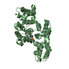
| Entry | Database: PDB / ID: 1mcx | ||||||
|---|---|---|---|---|---|---|---|
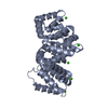
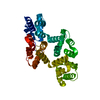
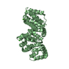
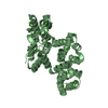
| Title | STRUCTURE OF FULL-LENGTH ANNEXIN A1 IN THE PRESENCE OF CALCIUM | ||||||
 Components Components | ANNEXIN I | ||||||
 Keywords Keywords | METAL BINDING PROTEIN / calcium/phospholipid binding protein | ||||||
| Function / homology |  Function and homology information Function and homology informationregulation of interleukin-1 production / granulocyte chemotaxis / regulation of leukocyte migration / positive regulation of T-helper 1 cell differentiation / phospholipase A2 inhibitor activity / regulation of hormone secretion / neutrophil activation / calcium-dependent phospholipid binding / negative regulation of T-helper 2 cell differentiation / motile cilium ...regulation of interleukin-1 production / granulocyte chemotaxis / regulation of leukocyte migration / positive regulation of T-helper 1 cell differentiation / phospholipase A2 inhibitor activity / regulation of hormone secretion / neutrophil activation / calcium-dependent phospholipid binding / negative regulation of T-helper 2 cell differentiation / motile cilium / vesicle membrane / cellular response to glucocorticoid stimulus / negative regulation of exocytosis / phosphatidylserine binding / positive regulation of wound healing / monocyte chemotaxis / lateral plasma membrane / G protein-coupled receptor signaling pathway, coupled to cyclic nucleotide second messenger / positive regulation of interleukin-2 production / phagocytosis / phagocytic cup / positive regulation of T cell proliferation / regulation of cell shape / actin cytoskeleton organization / regulation of inflammatory response / early endosome membrane / basolateral plasma membrane / adaptive immune response / apical plasma membrane / inflammatory response / innate immune response / calcium ion binding / signal transduction / : / extracellular exosome / nucleus / plasma membrane / cytoplasm Similarity search - Function | ||||||
| Biological species |  | ||||||
| Method |  X-RAY DIFFRACTION / SYNCHROTRON / MOLECULAR REPLACEMENT / Resolution: 2.03 Å X-RAY DIFFRACTION / SYNCHROTRON / MOLECULAR REPLACEMENT / Resolution: 2.03 Å | ||||||
 Authors Authors | Luecke, H. / Rosengarth, A. | ||||||
 Citation Citation | Journal: J.Mol.Biol. / Year: 2003 Title: A Calcium-driven Conformational Switch of the N-terminaland Core Domains of Annexin A1 Authors: Luecke, H. / Rosengarth, A. | ||||||
| History |
|
- Structure visualization
| Structure viewer | Molecule: MolmilJmol/JSmol |
|---|
- Downloads & links
-Download
| PDBx/mmCIF format | 1mcx.cif.gz | 84.4 KB | Display | PDBx/mmCIF format |
|---|---|---|---|---|
| PDB format | pdb1mcx.ent.gz | 61.7 KB | Display | PDB format |
| PDBx/mmJSON format | 1mcx.json.gz | Tree view | PDBx/mmJSON format | |
| Others |  Other downloads Other downloads |
-Validation report
| Arichive directory | https://data.pdbj.org/pub/pdb/validation_reports/mc/1mcxftp://data.pdbj.org/pub/pdb/validation_reports/mc/1mcx | HTTPS FTP |
|---|
-Related structure data
| Related structure data |  1hm6S S: Starting model for refinement |
|---|---|
| Similar structure data |


-Links
 PDBj
PDBj- Assembly
| Deposited unit | 
| ||||||||
|---|---|---|---|---|---|---|---|---|---|
| 1 |
| ||||||||
| Unit cell |
|
-Components
| #1: Protein | Mass: 38802.355 Da / Num. of mol.: 1 / Mutation: S51L, H69L, L231P, N289I Source method: isolated from a genetically manipulated source Source: (gene. exp.)  | ||||
|---|---|---|---|---|---|
| #2: Chemical | ChemComp-CA /   Mass: 40.078 Da / Num. of mol.: 8 / Source method: obtained synthetically / Formula: Ca Mass: 40.078 Da / Num. of mol.: 8 / Source method: obtained synthetically / Formula: Ca#3: Water | ChemComp-HOH / |  Mass: 18.015 Da / Num. of mol.: 386 / Source method: isolated from a natural source / Formula: H2O Mass: 18.015 Da / Num. of mol.: 386 / Source method: isolated from a natural source / Formula: H2OHas protein modification | Y | |
-Experimental details
-Experiment
| Experiment | Method: X-RAY DIFFRACTION / Number of used crystals: 1 |
|---|
- Sample preparation
| Crystal | Density Matthews: 3.6 Å3/Da / Density % sol: 65.84 % | ||||||||||||||||||||||||
|---|---|---|---|---|---|---|---|---|---|---|---|---|---|---|---|---|---|---|---|---|---|---|---|---|---|
| Crystal grow | Temperature: 277 K / Method: vapor diffusion, hanging drop / pH: 8 Details: PEG 1000, imidazol, calciumacetate, pH 8.0, VAPOR DIFFUSION, HANGING DROP, temperature 277K | ||||||||||||||||||||||||
| Crystal grow | *PLUS Method: unknown | ||||||||||||||||||||||||
| Components of the solutions | *PLUS
|
-Data collection
| Diffraction | Mean temperature: 100 K |
|---|---|
| Diffraction source | Source: SYNCHROTRON / Site: ALS  / Beamline: 5.0.1 / Wavelength: 1 Å / Beamline: 5.0.1 / Wavelength: 1 Å |
| Detector | Type: ADSC QUANTUM 4 / Detector: CCD / Date: Mar 26, 2002 |
| Radiation | Protocol: SINGLE WAVELENGTH / Monochromatic (M) / Laue (L): M / Scattering type: x-ray |
| Radiation wavelength | Wavelength: 1 Å / Relative weight: 1 |
| Reflection | Resolution: 2.03→60 Å / Num. obs: 37069 / Observed criterion σ(I): -3 / Redundancy: 15.3 % / Rmerge(I) obs: 0.117 / Net I/σ(I): 27.2 |
| Reflection | *PLUS Lowest resolution: 60 Å / % possible obs: 97.9 % / Num. measured all: 568454 |
| Reflection shell | *PLUS Highest resolution: 2.03 Å / Lowest resolution: 2.07 Å / % possible obs: 95.7 % / Rmerge(I) obs: 0.891 / Mean I/σ(I) obs: 3.3 |
- Processing
| Software |
| |||||||||||||||||||||||||
|---|---|---|---|---|---|---|---|---|---|---|---|---|---|---|---|---|---|---|---|---|---|---|---|---|---|---|
| Refinement | Method to determine structure: MOLECULAR REPLACEMENT Starting model: PDB ENTRY 1HM6 Resolution: 2.03→60 Å / Isotropic thermal model: isotropic / Cross valid method: Rfree / σ(F): 0 / σ(I): 0 / Stereochemistry target values: Engh & Huber
| |||||||||||||||||||||||||
| Displacement parameters | Biso mean: 26.7 Å2 | |||||||||||||||||||||||||
| Refinement step | Cycle: LAST / Resolution: 2.03→60 Å
| |||||||||||||||||||||||||
| Refine LS restraints |
| |||||||||||||||||||||||||
| Refinement | *PLUS Lowest resolution: 60 Å / Rfactor Rfree: 0.2311 / Rfactor Rwork: 0.1962 | |||||||||||||||||||||||||
| Solvent computation | *PLUS | |||||||||||||||||||||||||
| Displacement parameters | *PLUS | |||||||||||||||||||||||||
| Refine LS restraints | *PLUS Type: c_angle_deg / Dev ideal: 1.061 |
