 Movie
Movie Controller


+ Open data

- Basic information
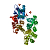
| Entry | Database: PDB / ID: 1hm6 | ||||||
|---|---|---|---|---|---|---|---|
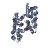
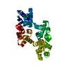
| Title | X-RAY STRUCTURE OF FULL-LENGTH ANNEXIN 1 | ||||||
 Components Components | ANNEXIN 1 | ||||||
 Keywords Keywords | METAL / LIPID BINDING PROTEIN / phospholipid/Ca(2+)-binding protein / calcium-free form / full-length protein comprising protein core and N-terminal domain | ||||||
| Function / homology |  Function and homology information Function and homology informationregulation of interleukin-1 production / granulocyte chemotaxis / regulation of leukocyte migration / positive regulation of T-helper 1 cell differentiation / phospholipase A2 inhibitor activity / regulation of hormone secretion / neutrophil activation / calcium-dependent phospholipid binding / negative regulation of T-helper 2 cell differentiation / motile cilium ...regulation of interleukin-1 production / granulocyte chemotaxis / regulation of leukocyte migration / positive regulation of T-helper 1 cell differentiation / phospholipase A2 inhibitor activity / regulation of hormone secretion / neutrophil activation / calcium-dependent phospholipid binding / negative regulation of T-helper 2 cell differentiation / motile cilium / vesicle membrane / cellular response to glucocorticoid stimulus / negative regulation of exocytosis / phosphatidylserine binding / positive regulation of wound healing / monocyte chemotaxis / lateral plasma membrane / G protein-coupled receptor signaling pathway, coupled to cyclic nucleotide second messenger / positive regulation of interleukin-2 production / phagocytosis / phagocytic cup / positive regulation of T cell proliferation / regulation of cell shape / actin cytoskeleton organization / regulation of inflammatory response / early endosome membrane / basolateral plasma membrane / adaptive immune response / apical plasma membrane / inflammatory response / innate immune response / calcium ion binding / signal transduction / : / extracellular exosome / nucleus / plasma membrane / cytoplasm Similarity search - Function | ||||||
| Biological species |  | ||||||
| Method |  X-RAY DIFFRACTION / SYNCHROTRON / MIR, MOLECULAR REPLACEMENT / Resolution: 1.8 Å X-RAY DIFFRACTION / SYNCHROTRON / MIR, MOLECULAR REPLACEMENT / Resolution: 1.8 Å | ||||||
 Authors Authors | Rosengarth, A. / Gerke, V. / Luecke, H. | ||||||
 Citation Citation | Journal: J.Mol.Biol. / Year: 2001 Title: X-ray structure of full-length annexin 1 and implications for membrane aggregation. Authors: Rosengarth, A. / Gerke, V. / Luecke, H. #1: Journal: Acta Crystallogr.,Sect.D / Year: 2000Title: Crystallization and Preliminary X-ray Analysis of Full-length Annexin I Comprising the Core and N-terminal Domain. Authors: Rosengarth, A. / Luecke, H. | ||||||
| History |
| ||||||
| Remark 300 | BIOMOLECULE: 1 THIS ENTRY CONTAINS THE CRYSTALLOGRAPHIC ASYMMETRIC UNIT WHICH CONSISTS OF 2 CHAIN(S) ...BIOMOLECULE: 1 THIS ENTRY CONTAINS THE CRYSTALLOGRAPHIC ASYMMETRIC UNIT WHICH CONSISTS OF 2 CHAIN(S). THE BIOLOGICAL MOLECULE MAY BE A MONOMER OR HIGHER OLIGOMER. | ||||||
| Remark 999 | SEQUENCE Seemann et al. deposited the cDNA sequence in genbank that they obtained from a library ... SEQUENCE Seemann et al. deposited the cDNA sequence in genbank that they obtained from a library screening. However, they were not able to get the complete cDNA. Later on, they introduced the naturally occuring (but missing) 5 amino acid residues at the N-terminal domain by PCR. This is also described in their paper (Seemann et al. (1996), Mol. Biol. Cell 7, 1359-1374). According to Seemann et al. (1996), the sequence shown herein starts with residue Met1, that would be residue -5 according to the truncated genbank entry, although there was no electron density observed for this residue. The amino acid sequence does not exactly match the (DNA) sequence deposited in the genbank by Seemann et al. However, the authors received the original construct from them and the amino acid sequence is according to his paper (Seemann et al., (1996), see above) (confirmed by amino acid sequencing). The electron densities of the respective amino acid residues in the annealed omit map showed clearly that the following changes have to be made in comparison to his genbank sequence: 1) residue 51 is a Leu (not Ser) 2) residue 69 is a Leu (not His) 3) residue 231 is a Pro (not Leu) 4) residue 289 is a Ile (not Asn) |
- Structure visualization
| Structure viewer | Molecule: MolmilJmol/JSmol |
|---|
- Downloads & links
-Download
| PDBx/mmCIF format | 1hm6.cif.gz | 162.7 KB | Display | PDBx/mmCIF format |
|---|---|---|---|---|
| PDB format | pdb1hm6.ent.gz | 127.8 KB | Display | PDB format |
| PDBx/mmJSON format | 1hm6.json.gz | Tree view | PDBx/mmJSON format | |
| Others |  Other downloads Other downloads |
-Validation report
| Arichive directory | https://data.pdbj.org/pub/pdb/validation_reports/hm/1hm6ftp://data.pdbj.org/pub/pdb/validation_reports/hm/1hm6 | HTTPS FTP |
|---|
-Related structure data
| Related structure data |  1ainS S: Starting model for refinement |
|---|---|
| Similar structure data |


-Links
 PDBj
PDBj- Assembly
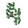
| Deposited unit | 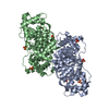
| ||||||||
|---|---|---|---|---|---|---|---|---|---|
| 1 | 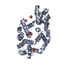
| ||||||||
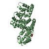
| 2 | 
| ||||||||
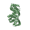
| Unit cell |
|
-Components
| #1: Protein | Mass: 38802.355 Da / Num. of mol.: 2 Source method: isolated from a genetically manipulated source Source: (gene. exp.)  #2: Chemical | ChemComp-SO4 /   Mass: 96.063 Da / Num. of mol.: 12 / Source method: obtained synthetically / Formula: SO4 Mass: 96.063 Da / Num. of mol.: 12 / Source method: obtained synthetically / Formula: SO4#3: Water | ChemComp-HOH / |  Mass: 18.015 Da / Num. of mol.: 739 / Source method: isolated from a natural source / Formula: H2O Mass: 18.015 Da / Num. of mol.: 739 / Source method: isolated from a natural source / Formula: H2OHas protein modification | Y | |
|---|
-Experimental details
-Experiment
| Experiment | Method: X-RAY DIFFRACTION / Number of used crystals: 1 |
|---|
- Sample preparation
| Crystal | Density Matthews: 2.52 Å3/Da / Density % sol: 51.08 % | ||||||||||||||||||||
|---|---|---|---|---|---|---|---|---|---|---|---|---|---|---|---|---|---|---|---|---|---|
| Crystal grow | Temperature: 277 K / Method: vapor diffusion, hanging drop / pH: 8.5 Details: 2.2 M Ammonium sulfate, 0.1 M Tris/HCl, pH 8.5, VAPOR DIFFUSION, HANGING DROP, temperature 277K | ||||||||||||||||||||
| Crystal grow | *PLUS Temperature: 4 ℃ / Method: vapor diffusion | ||||||||||||||||||||
| Components of the solutions | *PLUS
|
-Data collection
| Diffraction | Mean temperature: 100 K |
|---|---|
| Diffraction source | Source: SYNCHROTRON / Site: ALS  / Beamline: 5.0.2 / Wavelength: 1 Å / Beamline: 5.0.2 / Wavelength: 1 Å |
| Detector | Type: ADSC QUANTUM 4 / Detector: CCD / Date: Oct 28, 1999 |
| Radiation | Protocol: SINGLE WAVELENGTH / Monochromatic (M) / Laue (L): M / Scattering type: x-ray |
| Radiation wavelength | Wavelength: 1 Å / Relative weight: 1 |
| Reflection | Resolution: 1.8→40 Å / Num. all: 68225 / Num. obs: 68225 / % possible obs: 93 % / Redundancy: 10 % / Rmerge(I) obs: 0.082 / Net I/σ(I): 14.6 |
| Reflection shell | Resolution: 1.8→1.83 Å / Rmerge(I) obs: 0.599 / Mean I/σ(I) obs: 2.1 / Num. unique all: 3596 / % possible all: 99.4 |
| Reflection | *PLUS Num. measured all: 679717 |
| Reflection shell | *PLUS % possible obs: 99.4 % |
- Processing
| Software |
| ||||||||||||||||||||||||||||
|---|---|---|---|---|---|---|---|---|---|---|---|---|---|---|---|---|---|---|---|---|---|---|---|---|---|---|---|---|---|
| Refinement | Method to determine structure: MIR, MOLECULAR REPLACEMENT Starting model: PDB entry 1AIN Resolution: 1.8→40 Å / Cross valid method: RFREE / σ(F): 0 / σ(I): 0 / Stereochemistry target values: Engh & Huber
| ||||||||||||||||||||||||||||
| Displacement parameters | Biso mean: 26.62 Å2 | ||||||||||||||||||||||||||||
| Refinement step | Cycle: LAST / Resolution: 1.8→40 Å
| ||||||||||||||||||||||||||||
| Refine LS restraints |
| ||||||||||||||||||||||||||||
| Xplor file |
| ||||||||||||||||||||||||||||
| Software | *PLUS Name: CNS / Version: 1 / Classification: refinement | ||||||||||||||||||||||||||||
| Refinement | *PLUS Lowest resolution: 40 Å / σ(F): 0 / % reflection Rfree: 5 % / Rfactor all: 0.223 | ||||||||||||||||||||||||||||
| Solvent computation | *PLUS | ||||||||||||||||||||||||||||
| Displacement parameters | *PLUS | ||||||||||||||||||||||||||||
| Refine LS restraints | *PLUS
|
