 Movie
Movie Controller


+ Open data

- Basic information
| Entry | Database: PDB / ID: 1aii | |||||||||
|---|---|---|---|---|---|---|---|---|---|---|
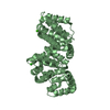
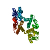
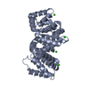
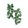
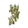
| Title | ANNEXIN III | |||||||||
 Components Components | ANNEXIN III | |||||||||
 Keywords Keywords | CALCIUM/PHOSPHOLIPID BINDING PROTEIN / ANNEXIN / PHOSPHOLIPASE A2 INHIBITOR / CALCIUM-PHOSPHOLIPID BINDING PROTEIN complex | |||||||||
| Function / homology |  Function and homology information Function and homology informationneutrophil degranulation / phospholipase A2 inhibitor activity / specific granule / calcium-dependent phospholipid binding / vesicle membrane / phosphatidylserine binding / : / phagocytosis / positive regulation of endothelial cell migration / Developmental Lineage of Pancreatic Ductal Cells ...neutrophil degranulation / phospholipase A2 inhibitor activity / specific granule / calcium-dependent phospholipid binding / vesicle membrane / phosphatidylserine binding / : / phagocytosis / positive regulation of endothelial cell migration / Developmental Lineage of Pancreatic Ductal Cells / phagocytic vesicle membrane / positive regulation of angiogenesis / calcium-dependent protein binding / defense response to bacterium / calcium ion binding / extracellular exosome / membrane / nucleus / plasma membrane / cytoplasm Similarity search - Function | |||||||||
| Biological species |  Homo sapiens (human) Homo sapiens (human) | |||||||||
| Method |  X-RAY DIFFRACTION / SYNCHROTRON / MOLECULAR REPLACEMENT / Resolution: 1.95 Å X-RAY DIFFRACTION / SYNCHROTRON / MOLECULAR REPLACEMENT / Resolution: 1.95 Å | |||||||||
 Authors Authors | Lewit-Bentley, A. / Perron, B. | |||||||||
 Citation Citation | Journal: J.Biol.Chem. / Year: 1997 Title: Can enzymatic activity, or otherwise, be inferred from structural studies of annexin III? Authors: Perron, B. / Lewit-Bentley, A. / Geny, B. / Russo-Marie, F. | |||||||||
| History |
|
- Structure visualization
| Structure viewer | Molecule: MolmilJmol/JSmol |
|---|
- Downloads & links
-Download
| PDBx/mmCIF format | 1aii.cif.gz | 83.8 KB | Display | PDBx/mmCIF format |
|---|---|---|---|---|
| PDB format | pdb1aii.ent.gz | 61.1 KB | Display | PDB format |
| PDBx/mmJSON format | 1aii.json.gz | Tree view | PDBx/mmJSON format | |
| Others |  Other downloads Other downloads |
-Validation report
| Arichive directory | https://data.pdbj.org/pub/pdb/validation_reports/ai/1aiiftp://data.pdbj.org/pub/pdb/validation_reports/ai/1aii | HTTPS FTP |
|---|
-Related structure data
| Related structure data |  1axnS S: Starting model for refinement |
|---|---|
| Similar structure data |


-Links
 PDBj
PDBj- Assembly
| Deposited unit | 
| ||||||||
|---|---|---|---|---|---|---|---|---|---|
| 1 |
| ||||||||
| Unit cell |
|
-Components
| #1: Protein | Mass: 36427.270 Da / Num. of mol.: 1 Source method: isolated from a genetically manipulated source Source: (gene. exp.) Homo sapiens (human)Description: PLASMID SOURCE PUC (BIOGEN, CAMBRIDGE, MA, USA) Plasmid: PGEX-2T / Production host:  | ||||||
|---|---|---|---|---|---|---|---|
| #2: Chemical | ChemComp-CA /   Mass: 40.078 Da / Num. of mol.: 5 / Source method: obtained synthetically / Formula: Ca Mass: 40.078 Da / Num. of mol.: 5 / Source method: obtained synthetically / Formula: Ca#3: Chemical | ChemComp-SO4 / |   Mass: 96.063 Da / Num. of mol.: 1 / Source method: obtained synthetically / Formula: SO4 Mass: 96.063 Da / Num. of mol.: 1 / Source method: obtained synthetically / Formula: SO4#4: Chemical | ChemComp-ETA / |   Type: L-peptide COOH carboxy terminus / Mass: 61.083 Da / Num. of mol.: 1 / Source method: obtained synthetically / Formula: C2H7NO Type: L-peptide COOH carboxy terminus / Mass: 61.083 Da / Num. of mol.: 1 / Source method: obtained synthetically / Formula: C2H7NO#5: Water | ChemComp-HOH / |  Mass: 18.015 Da / Num. of mol.: 233 / Source method: isolated from a natural source / Formula: H2O Mass: 18.015 Da / Num. of mol.: 233 / Source method: isolated from a natural source / Formula: H2O |
-Experimental details
-Experiment
| Experiment | Method: X-RAY DIFFRACTION / Number of used crystals: 1 |
|---|
- Sample preparation
| Crystal | Density Matthews: 2.22 Å3/Da / Density % sol: 38.87 % | ||||||||||||||||||||||||||||||||||||||||||||||||
|---|---|---|---|---|---|---|---|---|---|---|---|---|---|---|---|---|---|---|---|---|---|---|---|---|---|---|---|---|---|---|---|---|---|---|---|---|---|---|---|---|---|---|---|---|---|---|---|---|---|
| Crystal grow | pH: 7.8 Details: 18MG/ML PROTEIN, 10MM CACL2 IN 50 MM HEPES AT PH=7.8 WAS EQUILIBRATED AGAINST 50% AMMONIUM SULFATE IN THE SAME BUFFER. | ||||||||||||||||||||||||||||||||||||||||||||||||
| Crystal grow | *PLUS pH: 7.5 / Method: vapor diffusion | ||||||||||||||||||||||||||||||||||||||||||||||||
| Components of the solutions | *PLUS
|
-Data collection
| Diffraction | Mean temperature: 300 K |
|---|---|
| Diffraction source | Source: SYNCHROTRON / Site: LURE  / Beamline: D41A / Wavelength: 1.38 / Beamline: D41A / Wavelength: 1.38 |
| Detector | Type: MARRESEARCH / Detector: IMAGE PLATE / Date: Jun 8, 1995 |
| Radiation | Monochromator: GE(111) / Monochromatic (M) / Laue (L): M / Scattering type: x-ray |
| Radiation wavelength | Wavelength: 1.38 Å / Relative weight: 1 |
| Reflection | Resolution: 1.95→18.95 Å / Num. obs: 20818 / % possible obs: 98.5 % / Observed criterion σ(I): 0 / Redundancy: 2.9 % / Biso Wilson estimate: 0.6 Å2 / Rsym value: 0.072 / Net I/σ(I): 4.71 |
| Reflection shell | Resolution: 1.95→2 Å / Redundancy: 2.08 % / Mean I/σ(I) obs: 4.7 / Rsym value: 0.139 / % possible all: 83.5 |
| Reflection | *PLUS Num. measured all: 78671 / Rmerge(I) obs: 0.072 |
| Reflection shell | *PLUS % possible obs: 83.5 % / Rmerge(I) obs: 0.139 |
- Processing
| Software |
| ||||||||||||||||||||||||||||||||||||||||||||||||||||||||||||||||||||||||||||||||||||
|---|---|---|---|---|---|---|---|---|---|---|---|---|---|---|---|---|---|---|---|---|---|---|---|---|---|---|---|---|---|---|---|---|---|---|---|---|---|---|---|---|---|---|---|---|---|---|---|---|---|---|---|---|---|---|---|---|---|---|---|---|---|---|---|---|---|---|---|---|---|---|---|---|---|---|---|---|---|---|---|---|---|---|---|---|---|
| Refinement | Method to determine structure: MOLECULAR REPLACEMENT Starting model: PDB ENTRY 1AXN Resolution: 1.95→20 Å / Cross valid method: THROUGHOUT / σ(F): 1
| ||||||||||||||||||||||||||||||||||||||||||||||||||||||||||||||||||||||||||||||||||||
| Displacement parameters | Biso mean: 18.8 Å2 | ||||||||||||||||||||||||||||||||||||||||||||||||||||||||||||||||||||||||||||||||||||
| Refinement step | Cycle: LAST / Resolution: 1.95→20 Å
| ||||||||||||||||||||||||||||||||||||||||||||||||||||||||||||||||||||||||||||||||||||
| Refine LS restraints |
| ||||||||||||||||||||||||||||||||||||||||||||||||||||||||||||||||||||||||||||||||||||
| Software | *PLUS Name: PROLSQ / Classification: refinement | ||||||||||||||||||||||||||||||||||||||||||||||||||||||||||||||||||||||||||||||||||||
| Refinement | *PLUS Rfactor obs: 0.193 | ||||||||||||||||||||||||||||||||||||||||||||||||||||||||||||||||||||||||||||||||||||
| Solvent computation | *PLUS | ||||||||||||||||||||||||||||||||||||||||||||||||||||||||||||||||||||||||||||||||||||
| Displacement parameters | *PLUS | ||||||||||||||||||||||||||||||||||||||||||||||||||||||||||||||||||||||||||||||||||||
| Refine LS restraints | *PLUS
|
