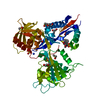
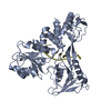
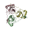
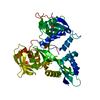
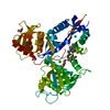
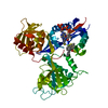
Entry Database : PDB / ID : 1a1vTitle HEPATITIS C VIRUS NS3 HELICASE DOMAIN COMPLEXED WITH SINGLE STRANDED SDNA DNA (5'-D(*UP*UP*UP*UP*UP*UP*UP*U)-3')PROTEIN (NS3 PROTEIN) Keywords / / / / / Function / homology Function Domain/homology Component
/ / / / / / / / / / / / / / / / / / / / / / / / / / / / / / / / / / / / / / / / / / / / / / / / / / / / / / / / / / / / / / / / / / / / / / / / / / / / / / / / / / / / / / / / / / / / / / / / / / / / / / / / / / / / / / / / / / / / / / / / / / / / / / / Biological species Method / / Resolution : 2.2 Å Authors Kim, J.L. / Morgenstern, K.A. / Griffith, J.P. / Dwyer, M.D. / Thomson, J.A. / Murcko, M.A. / Lin, C. / Caron, P.R. Journal : Structure / Year : 1998Title : Hepatitis C virus NS3 RNA helicase domain with a bound oligonucleotide: the crystal structure provides insights into the mode of unwinding.Authors : Kim, J.L. / Morgenstern, K.A. / Griffith, J.P. / Dwyer, M.D. / Thomson, J.A. / Murcko, M.A. / Lin, C. / Caron, P.R. History Deposition Dec 17, 1997 Processing site Revision 1.0 Jan 13, 1999 Provider / Type Revision 1.1 May 22, 2008 Group Revision 1.2 Jul 13, 2011 Group Revision 1.3 Oct 30, 2024 Group Data collection / Database references ... Data collection / Database references / Derived calculations / Structure summary Category chem_comp_atom / chem_comp_bond ... chem_comp_atom / chem_comp_bond / database_2 / pdbx_entry_details / pdbx_modification_feature / struct_conn / struct_ref_seq_dif / struct_site Item _database_2.pdbx_DOI / _database_2.pdbx_database_accession ... _database_2.pdbx_DOI / _database_2.pdbx_database_accession / _struct_conn.pdbx_leaving_atom_flag / _struct_conn.ptnr1_auth_comp_id / _struct_conn.ptnr1_auth_seq_id / _struct_conn.ptnr1_label_atom_id / _struct_conn.ptnr1_label_comp_id / _struct_conn.ptnr1_label_seq_id / _struct_conn.ptnr2_auth_comp_id / _struct_conn.ptnr2_auth_seq_id / _struct_conn.ptnr2_label_atom_id / _struct_conn.ptnr2_label_comp_id / _struct_conn.ptnr2_label_seq_id / _struct_ref_seq_dif.details / _struct_site.pdbx_auth_asym_id / _struct_site.pdbx_auth_comp_id / _struct_site.pdbx_auth_seq_id
Show all Show less
 Movie
Movie Controller

 Yorodumi
 Open data
 Basic information
 Components
 Keywords
 Function and homology information
 Hepatitis C virus
 X-RAY DIFFRACTION /
  Authors
 Citation
 Structure visualization
 Downloads & links
 Other downloads
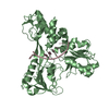

 PDBj
PDBj

 Assembly
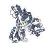


 Mass: 96.063 Da / Num. of mol.: 1 / Source method: obtained synthetically / Formula: SO4
 Mass: 18.015 Da / Num. of mol.: 159 / Source method: isolated from a natural source / Formula: H2O
 Sample preparation
 Processing