[English] 日本語
Yorodumi
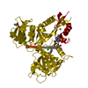
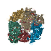
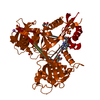
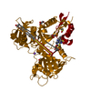
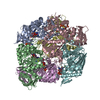
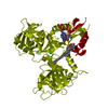
- PDB-5tvj: Crystal structure of acetyltransferase Eis from Mycobacterium tub... -

+
Open data


ID or keywords:

Loading...

-
Basic information

Entry
Database: PDB / ID: 5tvj
TitleCrystal structure of acetyltransferase Eis from Mycobacterium tuberculosis in complex with CoA and inhibitor 2k*: 1-(4-fluorophenyl)-2-[2-(4-methylphenyl)-2-oxoethyl]pyrrolo[1,2-a]pyrazin-2-ium
ComponentsEnhanced intracellular survival protein
KeywordsTRANSFERASE/TRANSFERASE Inhibitor / TRANSFERASE / acetyltransferase / resistance / aminoglycoside / TRANSFERASE-TRANSFERASE Inhibitor complex
Function / homology
Function and homology information


effector-mediated defense to host-produced reactive oxygen species / symbiont-mediated perturbation of host programmed cell death / symbiont-mediated perturbation of host inflammatory response / symbiont-mediated suppression of host defense-related programmed cell death / aminoglycoside antibiotic catabolic process / aminoglycoside N-acetyltransferase activity / symbiont-mediated perturbation of host innate immune response / Suppression of autophagy / bacterial extracellular vesicle / biological process involved in interaction with host ...effector-mediated defense to host-produced reactive oxygen species / symbiont-mediated perturbation of host programmed cell death / symbiont-mediated perturbation of host inflammatory response / symbiont-mediated suppression of host defense-related programmed cell death / aminoglycoside antibiotic catabolic process / aminoglycoside N-acetyltransferase activity / symbiont-mediated perturbation of host innate immune response / Suppression of autophagy / bacterial extracellular vesicle / biological process involved in interaction with host / host cell cytoplasmic vesicle / N-acetyltransferase activity / peptide-lysine-N-acetyltransferase activity / Transferases; Acyltransferases; Transferring groups other than aminoacyl groups / host extracellular space / response to antibiotic / identical protein binding / cytosol
Similarity search - Function
N-acetyltransferase Eis / Enhanced intracellular survival protein domain / Eis-like, acetyltransferase domain / Sterol carrier protein domain / Acetyltransferase (GNAT) domain / Acetyltransferase (GNAT) domain / SCP2 sterol-binding domain / Nonspecific Lipid-transfer Protein; Chain A / SCP2 sterol-binding domain superfamily / Gcn5-related N-acetyltransferase (GNAT) ...N-acetyltransferase Eis / Enhanced intracellular survival protein domain / Eis-like, acetyltransferase domain / Sterol carrier protein domain / Acetyltransferase (GNAT) domain / Acetyltransferase (GNAT) domain / SCP2 sterol-binding domain / Nonspecific Lipid-transfer Protein; Chain A / SCP2 sterol-binding domain superfamily / Gcn5-related N-acetyltransferase (GNAT) / Gcn5-related N-acetyltransferase (GNAT) domain profile. / GNAT domain / Acyl-CoA N-acyltransferase / Aminopeptidase / 2-Layer Sandwich / 3-Layer(aba) Sandwich / Alpha Beta
Similarity search - Domain/homology
Chem-7LF / COENZYME A / N-acetyltransferase Eis
Similarity search - Component
Biological speciesMycobacterium tuberculosis (bacteria)
MethodX-RAY DIFFRACTION / SYNCHROTRON / MOLECULAR REPLACEMENT / Resolution: 2.3 Å
AuthorsGajadeera, C.S. / Garzan, A. / Hou, C. / Garneau-Tsodikova, S. / Tsodikov, O.V.
Funding support United States, 1items
OrganizationGrant numberCountry
National Institutes of Health/National Institute Of Allergy and Infectious Diseases (NIH/NIAID)AI090048 United States
CitationJournal: ACS Infect Dis / Year: 2017
Title: Combating Enhanced Intracellular Survival (Eis)-Mediated Kanamycin Resistance of Mycobacterium tuberculosis by Novel Pyrrolo[1,5-a]pyrazine-Based Eis Inhibitors.
Authors: Garzan, A. / Willby, M.J. / Ngo, H.X. / Gajadeera, C.S. / Green, K.D. / Holbrook, S.Y. / Hou, C. / Posey, J.E. / Tsodikov, O.V. / Garneau-Tsodikova, S.
History
DepositionNov 9, 2016Deposition site: RCSB / Processing site: RCSB
Revision 1.0Mar 1, 2017Provider: repository / Type: Initial release
Revision 1.1Apr 26, 2017Group: Database references
Revision 1.2Sep 20, 2017Group: Author supporting evidence / Category: pdbx_audit_support / Item: _pdbx_audit_support.funding_organization
Revision 1.3Dec 11, 2019Group: Author supporting evidence / Category: pdbx_audit_support / Item: _pdbx_audit_support.funding_organization
Revision 1.4Oct 4, 2023Group: Data collection / Database references / Refinement description
Category: chem_comp_atom / chem_comp_bond ...chem_comp_atom / chem_comp_bond / database_2 / pdbx_initial_refinement_model
Item: _database_2.pdbx_DOI / _database_2.pdbx_database_accession

-
Structure visualization

Structure viewerMolecule:
MolmilJmol/JSmol

Downloads & links

-
Assembly

Deposited unit
A: Enhanced intracellular survival protein
hetero molecules


Theoretical massNumber of molelcules
Total (without water)47,2405
Polymers45,9991
Non-polymers1,2404
Water1,60389
1
A: Enhanced intracellular survival protein
hetero molecules
x 6


Theoretical massNumber of molelcules
Total (without water)283,43830
Polymers275,9956
Non-polymers7,44324
Water1086
TypeNameSymmetry operationNumber
identity operation1_555x,y,z1
crystal symmetry operation2_555-y,x-y,z1
crystal symmetry operation3_555-x+y,-x,z1
crystal symmetry operation4_556y,x,-z+11
crystal symmetry operation5_556x-y,-y,-z+11
crystal symmetry operation6_556-x,-x+y,-z+11
Buried area31280 Å2
ΔGint-106 kcal/mol
Surface area88330 Å2
MethodPISA
Unit cell
Length a, b, c (Å)175.106, 175.106, 122.940
Angle α, β, γ (deg.)90.00, 90.00, 120.00
Int Tables number155
Space group name H-MH32

-
Components

-
Protein , 1 types, 1 molecules A

#1: Protein Enhanced intracellular survival protein


Mass: 45999.113 Da / Num. of mol.: 1 / Mutation: C204A
Source method: isolated from a genetically manipulated source
Source: (gene. exp.) Mycobacterium tuberculosis (bacteria) / Strain: ATCC 25618 / H37Rv / Gene: eis, Rv2416c, MTCY253.04 / Production host: Escherichia coli (E. coli) / Strain (production host): BL21(DE3) / References: UniProt: P9WFK7

-
Non-polymers , 5 types, 93 molecules

#2: Chemical ChemComp-COA / COENZYME A / Coenzyme A


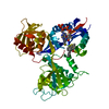
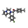
Mass: 767.534 Da / Num. of mol.: 1 / Source method: obtained synthetically / Formula: C21H36N7O16P3S
#3: Chemical ChemComp-7LF / 1-(4-fluorophenyl)-2-[2-(4-methylphenyl)-2-oxoethyl]pyrrolo[1,2-a]pyrazin-2-ium


Mass: 345.390 Da / Num. of mol.: 1 / Source method: obtained synthetically / Formula: C22H18FN2O
#4: Chemical ChemComp-GOL / GLYCEROL / GLYCERIN / PROPANE-1,2,3-TRIOL / Glycerol


Mass: 92.094 Da / Num. of mol.: 1 / Source method: obtained synthetically / Formula: C3H8O3
#5: Chemical ChemComp-CL / CHLORIDE ION / Chloride


Mass: 35.453 Da / Num. of mol.: 1 / Source method: obtained synthetically / Formula: Cl
#6: Water ChemComp-HOH / water / Water


Mass: 18.015 Da / Num. of mol.: 89 / Source method: isolated from a natural source / Formula: H2O

-
Experimental details

-
Experiment

ExperimentMethod: X-RAY DIFFRACTION / Number of used crystals: 1

-
Sample preparation

CrystalDensity Matthews: 3.94 Å3/Da / Density % sol: 68.8 %
Crystal growTemperature: 294 K / Method: vapor diffusion, hanging drop
Details: RESERVOIR: TRIS-HCL PH 8.5 ADJUSTED AT REMARK 280 ROOM TEMPERATURE (100 MM), PEG 8,000 (10-15% W/V), AND (NH4)2SO4 REMARK 280 (0.4 M), VAPOR DIFFUSION, HANGING DROP, TEMPERATURE 295K

-
Data collection

DiffractionMean temperature: 100 K
Diffraction sourceSource: SYNCHROTRON / Site: APS / Beamline: 22-ID / Wavelength: 1 Å
DetectorType: MARMOSAIC 300 mm CCD / Detector: CCD / Date: Jun 19, 2014
RadiationProtocol: SINGLE WAVELENGTH / Monochromatic (M) / Laue (L): M / Scattering type: x-ray
Radiation wavelengthWavelength: 1 Å / Relative weight: 1
ReflectionResolution: 2.3→40 Å / Num. obs: 30515 / % possible obs: 99.8 % / Redundancy: 7.3 % / Net I/σ(I): 24

-
Processing

Software
NameVersionClassification
REFMAC5.8.0071refinement
HKL-2000data reduction
HKL-2000data scaling
PHASERphasing
RefinementMethod to determine structure: MOLECULAR REPLACEMENT
Starting model: 3R1K
Resolution: 2.3→40 Å / Cor.coef. Fo:Fc: 0.958 / Cor.coef. Fo:Fc free: 0.944 / SU B: 6.191 / SU ML: 0.143 / Cross valid method: THROUGHOUT / ESU R: 0.192 / ESU R Free: 0.17 / Details: HYDROGENS HAVE BEEN ADDED IN THE RIDING POSITIONS
RfactorNum. reflection% reflectionSelection details
Rfree0.22365 1615 5 %RANDOM
Rwork0.19183 ---
obs0.19342 30515 99.8 %-
Solvent computationIon probe radii: 0.8 Å / Shrinkage radii: 0.8 Å / VDW probe radii: 1.2 Å
Displacement parametersBiso mean: 49.855 Å2
Baniso -1Baniso -2Baniso -3
1--2.25 Å2-1.13 Å20 Å2
2---2.25 Å20 Å2
3---7.31 Å2
Refinement stepCycle: 1 / Resolution: 2.3→40 Å
ProteinNucleic acidLigandSolventTotal
Num. atoms3047 0 81 89 3217
Refine LS restraints
Refine-IDTypeDev idealDev ideal targetNumber
X-RAY DIFFRACTIONr_bond_refined_d0.0070.0193199
X-RAY DIFFRACTIONr_bond_other_d0.0010.023022
X-RAY DIFFRACTIONr_angle_refined_deg1.4241.994357
X-RAY DIFFRACTIONr_angle_other_deg0.88336900
X-RAY DIFFRACTIONr_dihedral_angle_1_deg5.8225394
X-RAY DIFFRACTIONr_dihedral_angle_2_deg31.08421.348141
X-RAY DIFFRACTIONr_dihedral_angle_3_deg17.50515481
X-RAY DIFFRACTIONr_dihedral_angle_4_deg17.8231540
X-RAY DIFFRACTIONr_chiral_restr0.1780.2481
X-RAY DIFFRACTIONr_gen_planes_refined0.0060.0213606
X-RAY DIFFRACTIONr_gen_planes_other0.0040.02770
X-RAY DIFFRACTIONr_nbd_refined
X-RAY DIFFRACTIONr_nbd_other
X-RAY DIFFRACTIONr_nbtor_refined
X-RAY DIFFRACTIONr_nbtor_other
X-RAY DIFFRACTIONr_xyhbond_nbd_refined
X-RAY DIFFRACTIONr_xyhbond_nbd_other
X-RAY DIFFRACTIONr_metal_ion_refined
X-RAY DIFFRACTIONr_metal_ion_other
X-RAY DIFFRACTIONr_symmetry_vdw_refined
X-RAY DIFFRACTIONr_symmetry_vdw_other
X-RAY DIFFRACTIONr_symmetry_hbond_refined
X-RAY DIFFRACTIONr_symmetry_hbond_other
X-RAY DIFFRACTIONr_symmetry_metal_ion_refined
X-RAY DIFFRACTIONr_symmetry_metal_ion_other
X-RAY DIFFRACTIONr_mcbond_it2.2824.7521583
X-RAY DIFFRACTIONr_mcbond_other2.2824.7521581
X-RAY DIFFRACTIONr_mcangle_it3.7927.1161975
X-RAY DIFFRACTIONr_mcangle_other3.7927.1161976
X-RAY DIFFRACTIONr_scbond_it2.755.281616
X-RAY DIFFRACTIONr_scbond_other2.7445.281616
X-RAY DIFFRACTIONr_scangle_it
X-RAY DIFFRACTIONr_scangle_other4.5397.7762383
X-RAY DIFFRACTIONr_long_range_B_refined7.20839.0863446
X-RAY DIFFRACTIONr_long_range_B_other7.239.0573438
X-RAY DIFFRACTIONr_rigid_bond_restr
X-RAY DIFFRACTIONr_sphericity_free
X-RAY DIFFRACTIONr_sphericity_bonded
LS refinement shellResolution: 2.3→2.36 Å / Total num. of bins used: 20
RfactorNum. reflection% reflection
Rfree0.346 117 -
Rwork0.277 2208 -
obs--98.31 %

+
About Yorodumi

-
News

-
Feb 9, 2022. New format data for meta-information of EMDB entries

New format data for meta-information of EMDB entries

  • Version 3 of the EMDB header file is now the official format.
  • The previous official version 1.9 will be removed from the archive.

Related info.:EMDB header

External links:wwPDB to switch to version 3 of the EMDB data model

-
Aug 12, 2020. Covid-19 info

Covid-19 info

URL: https://pdbj.org/emnavi/covid19.php

New page: Covid-19 featured information page in EM Navigator.

Related info.:Covid-19 info / Mar 5, 2020. Novel coronavirus structure data

+
Mar 5, 2020. Novel coronavirus structure data

Novel coronavirus structure data

Related info.:Yorodumi Speices / Aug 12, 2020. Covid-19 info

External links:COVID-19 featured content - PDBj / Molecule of the Month (242):Coronavirus Proteases

+
Jan 31, 2019. EMDB accession codes are about to change! (news from PDBe EMDB page)

EMDB accession codes are about to change! (news from PDBe EMDB page)

  • The allocation of 4 digits for EMDB accession codes will soon come to an end. Whilst these codes will remain in use, new EMDB accession codes will include an additional digit and will expand incrementally as the available range of codes is exhausted. The current 4-digit format prefixed with “EMD-” (i.e. EMD-XXXX) will advance to a 5-digit format (i.e. EMD-XXXXX), and so on. It is currently estimated that the 4-digit codes will be depleted around Spring 2019, at which point the 5-digit format will come into force.
  • The EM Navigator/Yorodumi systems omit the EMD- prefix.

Related info.:Q: What is EMD? / ID/Accession-code notation in Yorodumi/EM Navigator

External links:EMDB Accession Codes are Changing Soon! / Contact to PDBj

+
Jul 12, 2017. Major update of PDB

Major update of PDB

  • wwPDB released updated PDB data conforming to the new PDBx/mmCIF dictionary.
  • This is a major update changing the version number from 4 to 5, and with Remediation, in which all the entries are updated.
  • In this update, many items about electron microscopy experimental information are reorganized (e.g. em_software).
  • Now, EM Navigator and Yorodumi are based on the updated data.

External links:wwPDB Remediation / Enriched Model Files Conforming to OneDep Data Standards Now Available in the PDB FTP Archive

-
Yorodumi

Thousand views of thousand structures

  • Yorodumi is a browser for structure data from EMDB, PDB, SASBDB, etc.
  • This page is also the successor to EM Navigator detail page, and also detail information page/front-end page for Omokage search.
  • The word "yorodu" (or yorozu) is an old Japanese word meaning "ten thousand". "mi" (miru) is to see.

Related info.:EMDB / PDB / SASBDB / Comparison of 3 databanks / Yorodumi Search / Aug 31, 2016. New EM Navigator & Yorodumi / Yorodumi Papers / Jmol/JSmol / Function and homology information / Changes in new EM Navigator and Yorodumi

Read more