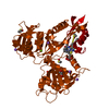
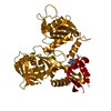
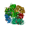
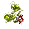
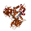
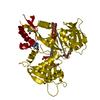
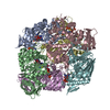
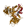
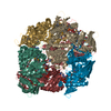
Crystal Structure of Enhanced Intracellular Survival (Eis) Protein from Mycobacterium tuberculosis with Acetyl CoA
Components
Enhanced intracellular survival protein
Keywords
TRANSFERASE / GNAT / Acetyltransferase
Function / homology
Function and homology information
effector-mediated defense to host-produced reactive oxygen species / symbiont-mediated perturbation of host inflammatory response / symbiont-mediated perturbation of host innate immune response / symbiont-mediated suppression of host programmed cell death / Suppression of autophagy / aminoglycoside antibiotic catabolic process / aminoglycoside N-acetyltransferase activity / bacterial extracellular vesicle / symbiont-mediated perturbation of host programmed cell death / biological process involved in interaction with host ...effector-mediated defense to host-produced reactive oxygen species / symbiont-mediated perturbation of host inflammatory response / symbiont-mediated perturbation of host innate immune response / symbiont-mediated suppression of host programmed cell death / Suppression of autophagy / aminoglycoside antibiotic catabolic process / aminoglycoside N-acetyltransferase activity / bacterial extracellular vesicle / symbiont-mediated perturbation of host programmed cell death / biological process involved in interaction with host / N-acetyltransferase activity / host cell cytoplasmic vesicle / protein-lysine-acetyltransferase activity / Transferases; Acyltransferases; Transferring groups other than aminoacyl groups / host extracellular region / response to antibiotic / identical protein binding / cytosol Similarity search - Function
In the structure databanks used in Yorodumi, some data are registered as the other names, "COVID-19 virus" and "2019-nCoV". Here are the details of the virus and the list of structure data.
Jan 31, 2019. EMDB accession codes are about to change! (news from PDBe EMDB page)
EMDB accession codes are about to change! (news from PDBe EMDB page)
The allocation of 4 digits for EMDB accession codes will soon come to an end. Whilst these codes will remain in use, new EMDB accession codes will include an additional digit and will expand incrementally as the available range of codes is exhausted. The current 4-digit format prefixed with “EMD-” (i.e. EMD-XXXX) will advance to a 5-digit format (i.e. EMD-XXXXX), and so on. It is currently estimated that the 4-digit codes will be depleted around Spring 2019, at which point the 5-digit format will come into force.
The EM Navigator/Yorodumi systems omit the EMD- prefix.
Related info.:Q: What is EMD? / ID/Accession-code notation in Yorodumi/EM Navigator
Yorodumi is a browser for structure data from EMDB, PDB, SASBDB, etc.
This page is also the successor to EM Navigator detail page, and also detail information page/front-end page for Omokage search.
The word "yorodu" (or yorozu) is an old Japanese word meaning "ten thousand". "mi" (miru) is to see.
Related info.:EMDB / PDB / SASBDB / Comparison of 3 databanks / Yorodumi Search / Aug 31, 2016. New EM Navigator & Yorodumi / Yorodumi Papers / Jmol/JSmol / Function and homology information / Changes in new EM Navigator and Yorodumi
 Movie
Movie Controller

 Yorodumi
 Open data
 Basic information
 Components
 Keywords
 Function and homology information
 Mycobacterium tuberculosis (bacteria)
 X-RAY DIFFRACTION /
  Authors
 Citation
 Structure visualization
 Downloads & links
 Other downloads
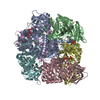


 PDBj
PDBj

 Assembly

 Mass: 809.571 Da / Num. of mol.: 12 / Source method: obtained synthetically / Formula: C23H38N7O17P3S
 Mass: 18.015 Da / Num. of mol.: 483 / Source method: isolated from a natural source / Formula: H2O
 Sample preparation
 / Beamline: BL-5A / Wavelength: 0.9792 Å
 Processing