 Movie
Movie Controller

[English] 日本語
 Yorodumi
- PDB-6f5h: Crystal structure of USP7 in complex with a 4-hydroxypiperidine b... -
+ Open data

- Basic information
| Entry | Database: PDB / ID: 6f5h | ||||||
|---|---|---|---|---|---|---|---|
| Title | Crystal structure of USP7 in complex with a 4-hydroxypiperidine based inhibitor | ||||||
 Components Components | Ubiquitin carboxyl-terminal hydrolase 7 | ||||||
 Keywords Keywords |  HYDROLASE / USP7 / reversible / inhibitor / selective HYDROLASE / USP7 / reversible / inhibitor / selective | ||||||
| Function / homology |  Function and homology information Function and homology informationregulation of telomere capping / positive regulation of DNA demethylation / monoubiquitinated protein deubiquitination / regulation of retrograde transport, endosome to Golgi / deubiquitinase activity / negative regulation of gene expression via chromosomal CpG island methylation / regulation of DNA-binding transcription factor activity / K48-linked deubiquitinase activity / symbiont-mediated disruption of host cell PML body / negative regulation of NF-kappaB transcription factor activity ...regulation of telomere capping / positive regulation of DNA demethylation / monoubiquitinated protein deubiquitination / regulation of retrograde transport, endosome to Golgi / deubiquitinase activity / negative regulation of gene expression via chromosomal CpG island methylation / regulation of DNA-binding transcription factor activity / K48-linked deubiquitinase activity / symbiont-mediated disruption of host cell PML body / negative regulation of NF-kappaB transcription factor activity / protein deubiquitination / negative regulation of proteasomal ubiquitin-dependent protein catabolic process / transcription-coupled nucleotide-excision repair / negative regulation of gluconeogenesis / negative regulation of TORC1 signaling / Regulation of PTEN localization / Synthesis of active ubiquitin: roles of E1 and E2 enzymes / regulation of signal transduction by p53 class mediator / regulation of protein stability / regulation of circadian rhythm / PML body / Transcription-Coupled Nucleotide Excision Repair (TC-NER) / Formation of TC-NER Pre-Incision Complex / Dual incision in TC-NER / Gap-filling DNA repair synthesis and ligation in TC-NER / rhythmic process / Regulation of TP53 Degradation / p53 binding / chromosome / ubiquitinyl hydrolase 1 / cysteine-type deubiquitinase activity / protein stabilization / protein ubiquitination / nuclear body / Ub-specific processing proteases / cysteine-type endopeptidase activity / protein-containing complex / proteolysis / nucleoplasm / nucleus / cytosolSimilarity search - Function | ||||||
| Biological species |  Homo sapiens (human) Homo sapiens (human) | ||||||
| Method | X-RAY DIFFRACTION / SYNCHROTRON / Resolution: 2.16 Å | ||||||
 Authors Authors | Harrison, T. / Gavory, G. / O'Dowd, C. / Helm, M. / Flasz, J. / Dossang, A. / Hughes, C. / Cassidy, E. / McClelland, K. / Odrzywol, E. ...Harrison, T. / Gavory, G. / O'Dowd, C. / Helm, M. / Flasz, J. / Dossang, A. / Hughes, C. / Cassidy, E. / McClelland, K. / Odrzywol, E. / Page, N. / Barker, O. / Miel, H. / Feutron-Burton, S. / Rountree, J.S.S. | ||||||
 Citation Citation | Journal: ACS Med Chem Lett / Year: 2018 Title: Identification and Structure-Guided Development of Pyrimidinone Based USP7 Inhibitors. Authors: O'Dowd, C.R. / Helm, M.D. / Rountree, J.S.S. / Flasz, J.T. / Arkoudis, E. / Miel, H. / Hewitt, P.R. / Jordan, L. / Barker, O. / Hughes, C. / Rozycka, E. / Cassidy, E. / McClelland, K. / ...Authors: O'Dowd, C.R. / Helm, M.D. / Rountree, J.S.S. / Flasz, J.T. / Arkoudis, E. / Miel, H. / Hewitt, P.R. / Jordan, L. / Barker, O. / Hughes, C. / Rozycka, E. / Cassidy, E. / McClelland, K. / Odrzywol, E. / Page, N. / Feutren-Burton, S. / Dvorkin, S. / Gavory, G. / Harrison, T. | ||||||
| History |
|
- Structure visualization
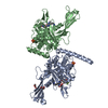
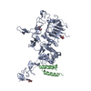
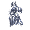
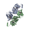
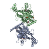
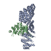
| Structure viewer | Molecule: MolmilJmol/JSmol |
|---|
- Downloads & links
-Download
| PDBx/mmCIF format | 6f5h.cif.gz | 296.3 KB | Display | PDBx/mmCIF format |
|---|---|---|---|---|
| PDB format | pdb6f5h.ent.gz | 248.6 KB | Display | PDB format |
| PDBx/mmJSON format | 6f5h.json.gz | Tree view | PDBx/mmJSON format | |
| Others |  Other downloads Other downloads |
-Validation report
| Arichive directory | https://data.pdbj.org/pub/pdb/validation_reports/f5/6f5hftp://data.pdbj.org/pub/pdb/validation_reports/f5/6f5h | HTTPS FTP |
|---|
-Related structure data
| Related structure data | |
|---|---|
| Similar structure data |


-Links
 PDBj
PDBj


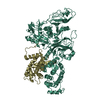
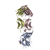
- Assembly
| Deposited unit | 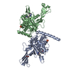
| ||||||||
|---|---|---|---|---|---|---|---|---|---|
| 1 |
| ||||||||
| Unit cell |
|
-Components
| #1: Protein | Mass: 41505.855 Da / Num. of mol.: 2 Source method: isolated from a genetically manipulated source Source: (gene. exp.) Homo sapiens (human) / Gene: USP7, HAUSP / Production host:  Escherichia coli (E. coli) / References: UniProt: Q93009, ubiquitinyl hydrolase 1 Escherichia coli (E. coli) / References: UniProt: Q93009, ubiquitinyl hydrolase 1#2: Chemical | Sulfate  Mass: 96.063 Da / Num. of mol.: 3 / Source method: obtained synthetically / Formula: SO4 Mass: 96.063 Da / Num. of mol.: 3 / Source method: obtained synthetically / Formula: SO4#3: Chemical | ChemComp-GOL / | Glycerol  Mass: 92.094 Da / Num. of mol.: 1 / Source method: obtained synthetically / Formula: C3H8O3 Mass: 92.094 Da / Num. of mol.: 1 / Source method: obtained synthetically / Formula: C3H8O3#4: Chemical | 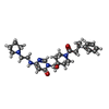  Mass: 467.604 Da / Num. of mol.: 2 / Source method: obtained synthetically / Formula: C26H37N5O3 / Feature type: SUBJECT OF INVESTIGATION Mass: 467.604 Da / Num. of mol.: 2 / Source method: obtained synthetically / Formula: C26H37N5O3 / Feature type: SUBJECT OF INVESTIGATION#5: Water | ChemComp-HOH / | Water Mass: 18.015 Da / Num. of mol.: 404 / Source method: isolated from a natural source / Formula: H2O Mass: 18.015 Da / Num. of mol.: 404 / Source method: isolated from a natural source / Formula: H2O |
|---|
-Experimental details
-Experiment
| Experiment | Method: X-RAY DIFFRACTION / Number of used crystals: 1 |
|---|
- Sample preparation
| Crystal | Density Matthews: 2.37 Å3/Da / Density % sol: 48 % |
|---|---|
| Crystal grow | Temperature: 293.15 K / Method: vapor diffusion, hanging drop / Details: PEG 4000, Tris, Li2-Sulfate, pH 7.75 |
-Data collection
| Diffraction | Mean temperature: 93 K |
|---|---|
| Diffraction source | Source: SYNCHROTRON / Site: ESRF  / Beamline: MASSIF-1 / Wavelength: 0.965 Å / Beamline: MASSIF-1 / Wavelength: 0.965 Å |
| Detector | Type: DECTRIS PILATUS3 2M / Detector: PIXEL / Date: Jan 27, 2015 |
| Radiation | Protocol: SINGLE WAVELENGTH / Monochromatic (M) / Laue (L): M / Scattering type: x-ray |
| Radiation wavelength | Wavelength: 0.965 Å / Relative weight: 1 |
| Reflection | Resolution: 2.16→29.95 Å / Num. obs: 39952 / % possible obs: 95.9 % / Redundancy: 2.4 % / Rrim(I) all: 0.11 / Net I/σ(I): 6.4 |
| Reflection shell | Resolution: 2.16→2.28 Å / Redundancy: 2.4 % / Mean I/σ(I) obs: 1.4 / Num. unique obs: 5872 / Rrim(I) all: 0.67 / % possible all: 96.8 |
- Processing
| Software |
| |||||||||||||||||||||||||||||||||||||||||||||||||||||||||||||||||||||||||||
|---|---|---|---|---|---|---|---|---|---|---|---|---|---|---|---|---|---|---|---|---|---|---|---|---|---|---|---|---|---|---|---|---|---|---|---|---|---|---|---|---|---|---|---|---|---|---|---|---|---|---|---|---|---|---|---|---|---|---|---|---|---|---|---|---|---|---|---|---|---|---|---|---|---|---|---|---|
| Refinement | Resolution: 2.16→29.95 Å / Cor.coef. Fo:Fc: 0.955 / Cor.coef. Fo:Fc free: 0.92 / SU B: 17.096 / SU ML: 0.221 / Cross valid method: THROUGHOUT / σ(F): 0 / ESU R: 0.304 / ESU R Free: 0.235 Details: HYDROGENS HAVE BEEN ADDED IN THE RIDING POSITIONS U VALUES : WITH TLS ADDED
| |||||||||||||||||||||||||||||||||||||||||||||||||||||||||||||||||||||||||||
| Solvent computation | Ion probe radii: 0.8 Å / Shrinkage radii: 0.8 Å / VDW probe radii: 1.2 Å | |||||||||||||||||||||||||||||||||||||||||||||||||||||||||||||||||||||||||||
| Displacement parameters | Biso max: 127.29 Å2 / Biso mean: 55.33 Å2 / Biso min: 32.01 Å2
| |||||||||||||||||||||||||||||||||||||||||||||||||||||||||||||||||||||||||||
| Refinement step | Cycle: final / Resolution: 2.16→29.95 Å
| |||||||||||||||||||||||||||||||||||||||||||||||||||||||||||||||||||||||||||
| Refine LS restraints |
| |||||||||||||||||||||||||||||||||||||||||||||||||||||||||||||||||||||||||||
| LS refinement shell | Resolution: 2.16→2.216 Å / Rfactor Rfree error: 0 / Total num. of bins used: 20
| |||||||||||||||||||||||||||||||||||||||||||||||||||||||||||||||||||||||||||
| Refinement TLS params. | Method: refined / Refine-ID: X-RAY DIFFRACTION
| |||||||||||||||||||||||||||||||||||||||||||||||||||||||||||||||||||||||||||
| Refinement TLS group |
|
