 Movie
Movie Controller

[English] 日本語
 Yorodumi
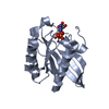
- PDB-5lnm: Crystal structure of D1050E mutant of the receiver domain of the ... -
+ Open data

- Basic information
| Entry | Database: PDB / ID: 5lnm | ||||||
|---|---|---|---|---|---|---|---|
| Title | Crystal structure of D1050E mutant of the receiver domain of the histidine kinase CKI1 from Arabidopsis thaliana | ||||||
 Components Components | Histidine kinase CKI1 | ||||||
 Keywords Keywords | TRANSFERASE / receiver domain / mutant / histidine kinase CKI1 / (alpha/beta)5 fold | ||||||
| Function / homology |  Function and homology information Function and homology informationsecondary growth / phloem or xylem histogenesis / embryo sac development / cytokinin-activated signaling pathway / protein histidine kinase activity / plasmodesma / phosphorelay sensor kinase activity / histidine kinase / protein homodimerization activity / plasma membrane Similarity search - Function | ||||||
| Biological species |  | ||||||
| Method |  X-RAY DIFFRACTION / SYNCHROTRON / MOLECULAR REPLACEMENT / Resolution: 1.95 Å X-RAY DIFFRACTION / SYNCHROTRON / MOLECULAR REPLACEMENT / Resolution: 1.95 Å | ||||||
 Authors Authors | Otrusinova, O. / Demo, G. / Kaderavek, P. / Jansen, S. / Jasenakova, Z. / Pekarova, B. / Janda, L. / Wimmerova, M. / Hejatko, J. / Zidek, L. | ||||||
 Citation Citation | Journal: J. Biol. Chem. / Year: 2017 Title: Conformational dynamics are a key factor in signaling mediated by the receiver domain of a sensor histidine kinase from Arabidopsis thaliana. Authors: Otrusinova, O. / Demo, G. / Padrta, P. / Jasenakova, Z. / Pekarova, B. / Gelova, Z. / Szmitkowska, A. / Kaderavek, P. / Jansen, S. / Zachrdla, M. / Klumpler, T. / Marek, J. / Hritz, J. / ...Authors: Otrusinova, O. / Demo, G. / Padrta, P. / Jasenakova, Z. / Pekarova, B. / Gelova, Z. / Szmitkowska, A. / Kaderavek, P. / Jansen, S. / Zachrdla, M. / Klumpler, T. / Marek, J. / Hritz, J. / Janda, L. / Iwai, H. / Wimmerova, M. / Hejatko, J. / Zidek, L. | ||||||
| History |
|
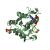
- Structure visualization
| Structure viewer | Molecule: MolmilJmol/JSmol |
|---|
- Downloads & links
-Download
| PDBx/mmCIF format | 5lnm.cif.gz | 47.4 KB | Display | PDBx/mmCIF format |
|---|---|---|---|---|
| PDB format | pdb5lnm.ent.gz | 32.3 KB | Display | PDB format |
| PDBx/mmJSON format | 5lnm.json.gz | Tree view | PDBx/mmJSON format | |
| Others |  Other downloads Other downloads |
-Validation report
| Arichive directory | https://data.pdbj.org/pub/pdb/validation_reports/ln/5lnmftp://data.pdbj.org/pub/pdb/validation_reports/ln/5lnm | HTTPS FTP |
|---|
-Related structure data
| Related structure data |  5lnnC  5n2nC  3mm4S S: Starting model for refinement C: citing same article ( |
|---|---|
| Similar structure data |


-Links
 PDBj
PDBj


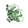
- Assembly
| Deposited unit | 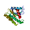
| ||||||||
|---|---|---|---|---|---|---|---|---|---|
| 1 |
| ||||||||
| Unit cell |
|
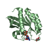
-Components
| #1: Protein | Mass: 23239.904 Da / Num. of mol.: 1 / Fragment: UNP residues 944-1122 Source method: isolated from a genetically manipulated source Source: (gene. exp.) Production host:  References: UniProt: O22267, histidine kinase |
|---|---|
| #2: Water | ChemComp-HOH /  Mass: 18.015 Da / Num. of mol.: 94 / Source method: isolated from a natural source / Formula: H2O Mass: 18.015 Da / Num. of mol.: 94 / Source method: isolated from a natural source / Formula: H2O |
-Experimental details
-Experiment
| Experiment | Method: X-RAY DIFFRACTION / Number of used crystals: 1 |
|---|
- Sample preparation
| Crystal | Density Matthews: 2.44 Å3/Da / Density % sol: 49.67 % |
|---|---|
| Crystal grow | Temperature: 290.15 K / Method: vapor diffusion, hanging drop / pH: 5.05 / Details: 2.54 M (NH4)2(SO4), 0.1 M MES pH 5.05 |
-Data collection
| Diffraction | Mean temperature: 100 K |
|---|---|
| Diffraction source | Source: SYNCHROTRON / Site: EMBL/DESY, HAMBURG  / Beamline: X12 / Wavelength: 0.97522 Å / Beamline: X12 / Wavelength: 0.97522 Å |
| Detector | Type: MARMOSAIC 225 mm CCD / Detector: CCD / Date: Nov 6, 2009 |
| Radiation | Monochromator: Si(111) / Protocol: SINGLE WAVELENGTH / Monochromatic (M) / Laue (L): M / Scattering type: x-ray |
| Radiation wavelength | Wavelength: 0.97522 Å / Relative weight: 1 |
| Reflection | Resolution: 1.95→47.6 Å / Num. obs: 15893 / % possible obs: 99.2 % / Redundancy: 5.7 % / Rmerge(I) obs: 0.117 / Net I/σ(I): 10.8 |
| Reflection shell | Resolution: 1.95→2.06 Å / Redundancy: 4 % / Rmerge(I) obs: 0.665 / Mean I/σ(I) obs: 1.6 / % possible all: 94.8 |
- Processing
| Software |
| ||||||||||||||||||||||||||||||||||||||||||||||||||||||||||||||||||||||||||||||||||||||||||||||||||||||||||||||||||||||||||||||||||||||||||||||||||||||||||||||||||||||||||||||||||||||
|---|---|---|---|---|---|---|---|---|---|---|---|---|---|---|---|---|---|---|---|---|---|---|---|---|---|---|---|---|---|---|---|---|---|---|---|---|---|---|---|---|---|---|---|---|---|---|---|---|---|---|---|---|---|---|---|---|---|---|---|---|---|---|---|---|---|---|---|---|---|---|---|---|---|---|---|---|---|---|---|---|---|---|---|---|---|---|---|---|---|---|---|---|---|---|---|---|---|---|---|---|---|---|---|---|---|---|---|---|---|---|---|---|---|---|---|---|---|---|---|---|---|---|---|---|---|---|---|---|---|---|---|---|---|---|---|---|---|---|---|---|---|---|---|---|---|---|---|---|---|---|---|---|---|---|---|---|---|---|---|---|---|---|---|---|---|---|---|---|---|---|---|---|---|---|---|---|---|---|---|---|---|---|---|
| Refinement | Method to determine structure: MOLECULAR REPLACEMENT Starting model: 3MM4 Resolution: 1.95→42.37 Å / Cor.coef. Fo:Fc: 0.94 / Cor.coef. Fo:Fc free: 0.934 / SU B: 3.96 / SU ML: 0.109 / Cross valid method: THROUGHOUT / ESU R: 0.174 / ESU R Free: 0.153 / Details: HYDROGENS HAVE BEEN ADDED IN THE RIDING POSITIONS
| ||||||||||||||||||||||||||||||||||||||||||||||||||||||||||||||||||||||||||||||||||||||||||||||||||||||||||||||||||||||||||||||||||||||||||||||||||||||||||||||||||||||||||||||||||||||
| Solvent computation | Ion probe radii: 0.8 Å / Shrinkage radii: 0.8 Å / VDW probe radii: 1.2 Å | ||||||||||||||||||||||||||||||||||||||||||||||||||||||||||||||||||||||||||||||||||||||||||||||||||||||||||||||||||||||||||||||||||||||||||||||||||||||||||||||||||||||||||||||||||||||
| Displacement parameters | Biso mean: 34.402 Å2
| ||||||||||||||||||||||||||||||||||||||||||||||||||||||||||||||||||||||||||||||||||||||||||||||||||||||||||||||||||||||||||||||||||||||||||||||||||||||||||||||||||||||||||||||||||||||
| Refinement step | Cycle: 1 / Resolution: 1.95→42.37 Å
| ||||||||||||||||||||||||||||||||||||||||||||||||||||||||||||||||||||||||||||||||||||||||||||||||||||||||||||||||||||||||||||||||||||||||||||||||||||||||||||||||||||||||||||||||||||||
| Refine LS restraints |
|