 Movie
Movie Controller


+ Open data

- Basic information
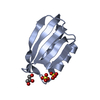
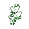
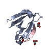
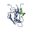
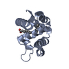
| Entry | Database: PDB / ID: 2ds2 | ||||||
|---|---|---|---|---|---|---|---|
| Title | Crystal structure of mabinlin II | ||||||
 Components Components |
| ||||||
 Keywords Keywords | PLANT PROTEIN / SEED STORAGE PROTEIN / SWEET PROTEIN | ||||||
| Function / homology |  Function and homology information Function and homology information | ||||||
| Biological species |  Capparis masaikai (plant) Capparis masaikai (plant) | ||||||
| Method |  X-RAY DIFFRACTION / SIRAS / Resolution: 1.7 Å X-RAY DIFFRACTION / SIRAS / Resolution: 1.7 Å | ||||||
 Authors Authors | Li, D.F. / Zhu, D.Y. / Wang, D.C. | ||||||
 Citation Citation | Journal: J.Struct.Biol. / Year: 2008 Title: Crystal structure of Mabinlin II: a novel structural type of sweet proteins and the main structural basis for its sweetness. Authors: Li, D.F. / Jiang, P. / Zhu, D.Y. / Hu, Y. / Max, M. / Wang, D.C. | ||||||
| History |
|
- Structure visualization
| Structure viewer | Molecule: MolmilJmol/JSmol |
|---|
- Downloads & links
-Download
| PDBx/mmCIF format | 2ds2.cif.gz | 47.9 KB | Display | PDBx/mmCIF format |
|---|---|---|---|---|
| PDB format | pdb2ds2.ent.gz | 38.4 KB | Display | PDB format |
| PDBx/mmJSON format | 2ds2.json.gz | Tree view | PDBx/mmJSON format | |
| Others |  Other downloads Other downloads |
-Validation report
| Summary document | 2ds2_validation.pdf.gz | 465.8 KB | Display | wwPDB validaton report |
|---|---|---|---|---|
| Full document | 2ds2_full_validation.pdf.gz | 473.7 KB | Display | |
| Data in XML | 2ds2_validation.xml.gz | 11.5 KB | Display | |
| Data in CIF | 2ds2_validation.cif.gz | 15 KB | Display | |
| Arichive directory | https://data.pdbj.org/pub/pdb/validation_reports/ds/2ds2ftp://data.pdbj.org/pub/pdb/validation_reports/ds/2ds2 | HTTPS FTP |
-Related structure data
| Similar structure data |
|---|


-Links
 PDBj
PDBj


- Assembly
| Deposited unit | 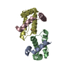
| ||||||||
|---|---|---|---|---|---|---|---|---|---|
| 1 | 
| ||||||||
| 2 | 
| ||||||||
| Unit cell |
|
-Components
| #1: Protein/peptide | Mass: 4174.806 Da / Num. of mol.: 2 / Source method: isolated from a natural source / Source: (natural) Capparis masaikai (plant) / Tissue: SEED / References: UniProt: P30233#2: Protein | Mass: 8327.779 Da / Num. of mol.: 2 / Source method: isolated from a natural source / Source: (natural) Capparis masaikai (plant) / Tissue: SEED / References: UniProt: P30233#3: Chemical | ChemComp-ACY / |   Mass: 60.052 Da / Num. of mol.: 1 / Source method: obtained synthetically / Formula: C2H4O2 Mass: 60.052 Da / Num. of mol.: 1 / Source method: obtained synthetically / Formula: C2H4O2#4: Water | ChemComp-HOH / |  Mass: 18.015 Da / Num. of mol.: 125 / Source method: isolated from a natural source / Formula: H2O Mass: 18.015 Da / Num. of mol.: 125 / Source method: isolated from a natural source / Formula: H2O |
|---|
-Experimental details
-Experiment
| Experiment | Method: X-RAY DIFFRACTION / Number of used crystals: 1 |
|---|
- Sample preparation
| Crystal | Density Matthews: 1.6 Å3/Da / Density % sol: 24.5 % |
|---|---|
| Crystal grow | Temperature: 310 K / Method: vapor diffusion, hanging drop / pH: 4.6 Details: SODIUM MALONATE, SODIUM ACETATE , pH 4.60, VAPOR DIFFUSION, HANGING DROP, temperature 310K |
-Data collection
| Diffraction | Mean temperature: 83 K |
|---|---|
| Diffraction source | Source: ROTATING ANODE / Type: RIGAKU / Wavelength: 1.5418 / Wavelength: 1.5418 Å |
| Detector | Type: RIGAKU RAXIS IV / Detector: IMAGE PLATE / Date: Apr 25, 2005 |
| Radiation | Monochromator: CON-FOCUSE / Protocol: SINGLE WAVELENGTH / Monochromatic (M) / Laue (L): M / Scattering type: x-ray |
| Radiation wavelength | Wavelength: 1.5418 Å / Relative weight: 1 |
| Reflection | Resolution: 1.7→20.5 Å / Num. obs: 17686 / % possible obs: 99.5 % / Observed criterion σ(F): 0 / Observed criterion σ(I): 0 / Redundancy: 3.6 % / Biso Wilson estimate: 30.7 Å2 / Rmerge(I) obs: 0.053 / Rsym value: 0.053 / Net I/σ(I): 6 |
| Reflection shell | Resolution: 1.7→1.79 Å / Redundancy: 3.5 % / Rmerge(I) obs: 0.266 / Mean I/σ(I) obs: 2.6 / Rsym value: 0.266 / % possible all: 100 |
- Processing
| Software |
| |||||||||||||||||||||||||
|---|---|---|---|---|---|---|---|---|---|---|---|---|---|---|---|---|---|---|---|---|---|---|---|---|---|---|
| Refinement | Method to determine structure: SIRAS / Resolution: 1.7→20.35 Å / σ(F): 0 / σ(I): 2 / Stereochemistry target values: Engh & Huber
| |||||||||||||||||||||||||
| Refine analyze |
| |||||||||||||||||||||||||
| Refinement step | Cycle: LAST / Resolution: 1.7→20.35 Å
| |||||||||||||||||||||||||
| Refine LS restraints |
| |||||||||||||||||||||||||
| LS refinement shell | Resolution: 1.7→1.81 Å / Rfactor Rfree error: 0.029
|
