 Movie
Movie Controller


+ Open data

- Basic information
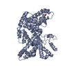
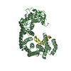
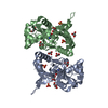
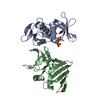
| Entry | Database: PDB / ID: 1mun | ||||||
|---|---|---|---|---|---|---|---|
| Title | CATALYTIC DOMAIN OF MUTY FROM ESCHERICHIA COLI D138N MUTANT | ||||||
 Components Components | ADENINE GLYCOSYLASE | ||||||
 Keywords Keywords |  DNA REPAIR / DNA G.A MISMATCH REPAIR ENZYME / GLYCOSIDASE / HYDROLASE DNA REPAIR / DNA G.A MISMATCH REPAIR ENZYME / GLYCOSIDASE / HYDROLASE | ||||||
| Function / homology |  Function and homology information Function and homology informationadenine/guanine mispair binding / adenine glycosylase / 8-oxo-7,8-dihydroguanine DNA N-glycosylase activity / purine-specific mismatch base pair DNA N-glycosylase activity / oxidized purine DNA binding / mismatch repair / base-excision repair / 4 iron, 4 sulfur cluster binding / metal ion bindingSimilarity search - Function | ||||||
| Biological species |  Escherichia coli (E. coli) Escherichia coli (E. coli) | ||||||
| Method | X-RAY DIFFRACTION / SYNCHROTRON / MOLECULAR REPLACEMENT / Resolution: 1.2 Å | ||||||
 Authors Authors | Guan, Y. / Tainer, J.A. | ||||||
 Citation Citation | Journal: Nat.Struct.Biol. / Year: 1998 Title: MutY catalytic core, mutant and bound adenine structures define specificity for DNA repair enzyme superfamily. Authors: Guan, Y. / Manuel, R.C. / Arvai, A.S. / Parikh, S.S. / Mol, C.D. / Miller, J.H. / Lloyd, S. / Tainer, J.A. | ||||||
| History |
|
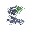
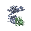
- Structure visualization
| Structure viewer | Molecule: MolmilJmol/JSmol |
|---|
- Downloads & links
-Download
| PDBx/mmCIF format | 1mun.cif.gz | 67.3 KB | Display | PDBx/mmCIF format |
|---|---|---|---|---|
| PDB format | pdb1mun.ent.gz | 48.3 KB | Display | PDB format |
| PDBx/mmJSON format | 1mun.json.gz | Tree view | PDBx/mmJSON format | |
| Others |  Other downloads Other downloads |
-Validation report
| Arichive directory | https://data.pdbj.org/pub/pdb/validation_reports/mu/1munftp://data.pdbj.org/pub/pdb/validation_reports/mu/1mun | HTTPS FTP |
|---|
-Related structure data


-Links
 PDBj
PDBj


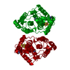
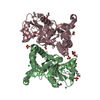
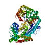
- Assembly
| Deposited unit | 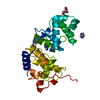
| |||||||||
|---|---|---|---|---|---|---|---|---|---|---|
| 1 | 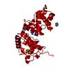
| |||||||||
| Unit cell |
| |||||||||
| Components on special symmetry positions |
|
-Components
| #1: Protein | Mass: 25048.004 Da / Num. of mol.: 1 / Fragment: CATALYTIC DOMAIN / Mutation: D138N Source method: isolated from a genetically manipulated source Source: (gene. exp.) Escherichia coli (E. coli)References: UniProt: P17802, Hydrolases; Glycosylases; Hydrolysing N-glycosyl compounds | ||||
|---|---|---|---|---|---|
| #2: Chemical | ChemComp-SF4 / Iron–sulfur cluster  Mass: 351.640 Da / Num. of mol.: 1 / Source method: obtained synthetically / Formula: Fe4S4 Mass: 351.640 Da / Num. of mol.: 1 / Source method: obtained synthetically / Formula: Fe4S4 | ||||
| #3: Chemical | ChemComp-IMD / Imidazole  Mass: 69.085 Da / Num. of mol.: 5 / Source method: obtained synthetically / Formula: C3H5N2 Mass: 69.085 Da / Num. of mol.: 5 / Source method: obtained synthetically / Formula: C3H5N2#4: Chemical | ChemComp-GOL / | Glycerol  Mass: 92.094 Da / Num. of mol.: 1 / Source method: obtained synthetically / Formula: C3H8O3 Mass: 92.094 Da / Num. of mol.: 1 / Source method: obtained synthetically / Formula: C3H8O3#5: Water | ChemComp-HOH / | Water Mass: 18.015 Da / Num. of mol.: 341 / Source method: isolated from a natural source / Formula: H2O Mass: 18.015 Da / Num. of mol.: 341 / Source method: isolated from a natural source / Formula: H2O |
-Experimental details
-Experiment
| Experiment | Method: X-RAY DIFFRACTION / Number of used crystals: 5 |
|---|
- Sample preparation
| Crystal | Density Matthews: 2.35 Å3/Da / Density % sol: 47.65 % | ||||||||||||||||||||||||||||||||||||
|---|---|---|---|---|---|---|---|---|---|---|---|---|---|---|---|---|---|---|---|---|---|---|---|---|---|---|---|---|---|---|---|---|---|---|---|---|---|
| Crystal grow | pH: 8 / Details: pH 8.0 | ||||||||||||||||||||||||||||||||||||
| Crystal grow | *PLUS Temperature: 15 ℃ / pH: 8.5 / Method: vapor diffusion, hanging drop | ||||||||||||||||||||||||||||||||||||
| Components of the solutions | *PLUS
|
-Data collection
| Diffraction | Mean temperature: 90 K |
|---|---|
| Diffraction source | Source: SYNCHROTRON / Site: SSRL  / Beamline: BL7-1 / Wavelength: 1.08 / Beamline: BL7-1 / Wavelength: 1.08 |
| Detector | Type: MARRESEARCH / Detector: IMAGE PLATE |
| Radiation | Monochromatic (M) / Laue (L): M / Scattering type: x-ray |
| Radiation wavelength | Wavelength: 1.08 Å / Relative weight: 1 |
| Reflection | Resolution: 1.2→20 Å / Num. obs: 65781 / % possible obs: 91 % / Observed criterion σ(I): 0 / Redundancy: 2.5 % / Rsym value: 0.053 |
| Reflection shell | Highest resolution: 1.2 Å |
| Reflection | *PLUS Num. measured all: 137211 / Rmerge(I) obs: 0.053 |
- Processing
| Software |
| |||||||||||||||||||||||||||||||||
|---|---|---|---|---|---|---|---|---|---|---|---|---|---|---|---|---|---|---|---|---|---|---|---|---|---|---|---|---|---|---|---|---|---|---|
| Refinement | Method to determine structure: MOLECULAR REPLACEMENT Starting model: NATIVE MUTY Resolution: 1.2→20 Å / Cross valid method: THROUGHOUT / σ(F): 0
| |||||||||||||||||||||||||||||||||
| Refine analyze | Num. disordered residues: 8 | |||||||||||||||||||||||||||||||||
| Refinement step | Cycle: LAST / Resolution: 1.2→20 Å
| |||||||||||||||||||||||||||||||||
| Refine LS restraints |
| |||||||||||||||||||||||||||||||||
| Software | *PLUS Name: SHELXL-97 / Classification: refinement | |||||||||||||||||||||||||||||||||
| Refinement | *PLUS % reflection Rfree: 5 % / Rfactor Rwork: 0.124 | |||||||||||||||||||||||||||||||||
| Solvent computation | *PLUS | |||||||||||||||||||||||||||||||||
| Displacement parameters | *PLUS |
