 Movie
Movie Controller

[English] 日本語
 Yorodumi
- PDB-6fof: Crystal structure of a crystallized variant of h-Gal3: Gal-3[NTS/... -
+ Open data

- Basic information
| Entry | Database: PDB / ID: 6fof | |||||||||
|---|---|---|---|---|---|---|---|---|---|---|
| Title | Crystal structure of a crystallized variant of h-Gal3: Gal-3[NTS/VII-IX] | |||||||||
 Components Components | Galectin-3,Galectin-3 | |||||||||
 Keywords Keywords | SUGAR BINDING PROTEIN / Galectin / apoptosis / glycosylation | |||||||||
| Function / homology |  Function and homology information Function and homology informationnegative regulation of NK T cell activation / negative regulation of immunological synapse formation / negative regulation of T cell activation via T cell receptor contact with antigen bound to MHC molecule on antigen presenting cell / disaccharide binding / RUNX2 regulates genes involved in differentiation of myeloid cells / regulation of T cell apoptotic process / mononuclear cell migration / receptor ligand inhibitor activity / negative regulation of endocytosis / positive regulation of mononuclear cell migration ...negative regulation of NK T cell activation / negative regulation of immunological synapse formation / negative regulation of T cell activation via T cell receptor contact with antigen bound to MHC molecule on antigen presenting cell / disaccharide binding / RUNX2 regulates genes involved in differentiation of myeloid cells / regulation of T cell apoptotic process / mononuclear cell migration / receptor ligand inhibitor activity / negative regulation of endocytosis / positive regulation of mononuclear cell migration / IgE binding / eosinophil chemotaxis / regulation of extrinsic apoptotic signaling pathway via death domain receptors / RUNX1 regulates transcription of genes involved in differentiation of myeloid cells / protein phosphatase inhibitor activity / positive chemotaxis / chemoattractant activity / positive regulation of calcium ion import / regulation of T cell proliferation / macrophage chemotaxis / Advanced glycosylation endproduct receptor signaling / monocyte chemotaxis / immunological synapse / ficolin-1-rich granule membrane / negative regulation of T cell receptor signaling pathway / neutrophil chemotaxis / epithelial cell differentiation / laminin binding / RNA splicing / secretory granule membrane / negative regulation of extrinsic apoptotic signaling pathway / positive regulation of protein localization to plasma membrane / spliceosomal complex / molecular condensate scaffold activity / positive regulation of protein-containing complex assembly / mRNA processing / carbohydrate binding / extracellular matrix / protein phosphatase binding / mitochondrial inner membrane / innate immune response / Neutrophil degranulation / cell surface / : / RNA binding / extracellular exosome / extracellular region / nucleoplasm / membrane / nucleus / plasma membrane / cytoplasm / cytosol Similarity search - Function | |||||||||
| Biological species |  Homo sapiens (human) Homo sapiens (human) | |||||||||
| Method |  X-RAY DIFFRACTION / SYNCHROTRON / MOLECULAR REPLACEMENT / Resolution: 2.2 Å X-RAY DIFFRACTION / SYNCHROTRON / MOLECULAR REPLACEMENT / Resolution: 2.2 Å | |||||||||
 Authors Authors | Romero, A. / Flores-Ibarra, A. / Medrano, F.J. | |||||||||
| Funding support |  Spain, 1items Spain, 1items
| |||||||||
 Citation Citation | Journal: Sci Rep / Year: 2018 Title: Crystallization of a human galectin-3 variant with two ordered segments in the shortened N-terminal tail. Authors: Flores-Ibarra, A. / Vertesy, S. / Medrano, F.J. / Gabius, H.J. / Romero, A. | |||||||||
| History |
|
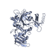
- Structure visualization
| Structure viewer | Molecule: MolmilJmol/JSmol |
|---|
- Downloads & links
-Download
| PDBx/mmCIF format | 6fof.cif.gz | 359.4 KB | Display | PDBx/mmCIF format |
|---|---|---|---|---|
| PDB format | pdb6fof.ent.gz | 291.2 KB | Display | PDB format |
| PDBx/mmJSON format | 6fof.json.gz | Tree view | PDBx/mmJSON format | |
| Others |  Other downloads Other downloads |
-Validation report
| Arichive directory | https://data.pdbj.org/pub/pdb/validation_reports/fo/6fofftp://data.pdbj.org/pub/pdb/validation_reports/fo/6fof | HTTPS FTP |
|---|
-Related structure data
| Related structure data | 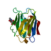 1a3kS S: Starting model for refinement |
|---|---|
| Similar structure data |


-Links
 PDBj
PDBj


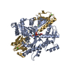
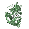
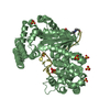
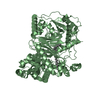
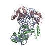
- Assembly
| Deposited unit | 
| ||||||||||||
|---|---|---|---|---|---|---|---|---|---|---|---|---|---|
| 1 | 
| ||||||||||||
| 2 | 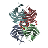
| ||||||||||||
| 3 | 
| ||||||||||||
| Unit cell |
|
-Components
| #1: Protein | Mass: 21209.848 Da / Num. of mol.: 12 Source method: isolated from a genetically manipulated source Source: (gene. exp.) Homo sapiens (human) / Gene: LGALS3, MAC2 / Production host:  #2: Polysaccharide | beta-D-galactopyranose-(1-4)-beta-D-glucopyranose / beta-lactose   Source method: isolated from a genetically manipulated source Details: oligosaccharide / References: beta-lactose #3: Chemical | ChemComp-SO4 /   Mass: 96.063 Da / Num. of mol.: 24 / Source method: obtained synthetically / Formula: SO4 Mass: 96.063 Da / Num. of mol.: 24 / Source method: obtained synthetically / Formula: SO4#4: Water | ChemComp-HOH / |  Mass: 18.015 Da / Num. of mol.: 128 / Source method: isolated from a natural source / Formula: H2O Mass: 18.015 Da / Num. of mol.: 128 / Source method: isolated from a natural source / Formula: H2O |
|---|
-Experimental details
-Experiment
| Experiment | Method: X-RAY DIFFRACTION / Number of used crystals: 1 |
|---|
- Sample preparation
| Crystal | Density Matthews: 2.78 Å3/Da / Density % sol: 55.4 % |
|---|---|
| Crystal grow | Temperature: 295 K / Method: vapor diffusion, sitting drop / pH: 8.5 Details: 18% PEG 8K, 100 mM Tris-HCl (pH 8.5), 200 mM lithium sulfate |
-Data collection
| Diffraction | Mean temperature: 100 K |
|---|---|
| Diffraction source | Source: SYNCHROTRON / Site: ALBA / Beamline: XALOC / Wavelength: 0.9794 Å |
| Detector | Type: DECTRIS PILATUS3 6M / Detector: PIXEL / Date: May 19, 2016 |
| Radiation | Protocol: SINGLE WAVELENGTH / Monochromatic (M) / Laue (L): M / Scattering type: x-ray |
| Radiation wavelength | Wavelength: 0.9794 Å / Relative weight: 1 |
| Reflection | Resolution: 2.2→49.1 Å / Num. obs: 112248 / % possible obs: 100 % / Observed criterion σ(F): 0 / Observed criterion σ(I): 0 / Redundancy: 9 % / Biso Wilson estimate: 43.25 Å2 / CC1/2: 0.997 / Rmerge(I) obs: 0.134 / Rsym value: 0.142 / Net I/σ(I): 11.7 |
| Reflection shell | Resolution: 2.2→2.32 Å / Redundancy: 9.1 % / Rmerge(I) obs: 1.233 / Mean I/σ(I) obs: 1.8 / Num. unique obs: 16179 / CC1/2: 0.558 / Rsym value: 1.308 / % possible all: 100 |
- Processing
| Software |
| ||||||||||||||||||||
|---|---|---|---|---|---|---|---|---|---|---|---|---|---|---|---|---|---|---|---|---|---|
| Refinement | Method to determine structure: MOLECULAR REPLACEMENT Starting model: 1A3K Resolution: 2.2→49.1 Å / Cross valid method: FREE R-VALUE / σ(F): 0
| ||||||||||||||||||||
| Solvent computation | Ion probe radii: 0.8 Å / Shrinkage radii: 0.8 Å / VDW probe radii: 1.2 Å | ||||||||||||||||||||
| Displacement parameters | Biso mean: 46.8 Å2
| ||||||||||||||||||||
| Refinement step | Cycle: LAST / Resolution: 2.2→49.1 Å
| ||||||||||||||||||||
| LS refinement shell | Resolution: 2.2→2.26 Å
|
