 Movie
Movie Controller

[English] 日本語
 Yorodumi
- PDB-1gtz: Structure of STREPTOMYCES COELICOLOR TYPE II DEHYDROQUINASE R23A ... -
+ Open data

- Basic information
| Entry | Database: PDB / ID: 1gtz | ||||||
|---|---|---|---|---|---|---|---|
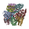
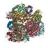
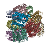
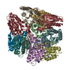
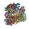
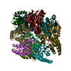
| Title | Structure of STREPTOMYCES COELICOLOR TYPE II DEHYDROQUINASE R23A MUTANT IN COMPLEX WITH DEHYDROSHIKIMATE | ||||||
 Components Components | 3-DEHYDROQUINATE DEHYDRATASE | ||||||
 Keywords Keywords | LYASE / TYPE II DEHYDROQUINASE / SHIKIMATE PATHWAY / DODECAMERIC QUATERNARY STRUCTURE / TETRAHEDRAL SYMMETRY AROMATIC AMINO ACID BIOSYNTHESIS | ||||||
| Function / homology |  Function and homology information Function and homology informationquinate catabolic process / 3-dehydroquinate dehydratase / 3-dehydroquinate dehydratase activity / chorismate biosynthetic process / aromatic amino acid family biosynthetic process / amino acid biosynthetic processSimilarity search - Function | ||||||
| Biological species |  STREPTOMYCES COELICOLOR (bacteria) STREPTOMYCES COELICOLOR (bacteria) | ||||||
| Method | X-RAY DIFFRACTION / SYNCHROTRON / MOLECULAR REPLACEMENT / Resolution: 1.6 Å | ||||||
 Authors Authors | Roszak, A.W. / Krell, T. / Robinson, D.A. / Hunter, I.S. / Coggins, J.R. / Lapthorn, A.J. | ||||||
 Citation Citation | Journal: Structure / Year: 2002 Title: The Structure and Mechanism of the Type II Dehydroquinase from Streptomyces Coelicolor Authors: Roszak, A.W. / Robinson, D.A. / Krell, T. / Hunter, I.S. / Fredrickson, M. / Abell, C. / Coggins, J.R. / Lapthorn, A.J. | ||||||
| History |
|
- Structure visualization
| Structure viewer | Molecule: MolmilJmol/JSmol |
|---|
- Downloads & links
-Download
| PDBx/mmCIF format | 1gtz.cif.gz | 721.6 KB | Display | PDBx/mmCIF format |
|---|---|---|---|---|
| PDB format | pdb1gtz.ent.gz | 600.7 KB | Display | PDB format |
| PDBx/mmJSON format | 1gtz.json.gz | Tree view | PDBx/mmJSON format | |
| Others |  Other downloads Other downloads |
-Validation report
| Arichive directory | https://data.pdbj.org/pub/pdb/validation_reports/gt/1gtzftp://data.pdbj.org/pub/pdb/validation_reports/gt/1gtz | HTTPS FTP |
|---|
-Related structure data
| Related structure data |  1d0iC  1gu0C  1gu1C  2dhqS C: citing same article ( S: Starting model for refinement |
|---|---|
| Similar structure data |


-Links
 PDBj
PDBj
- Assembly
| Deposited unit | 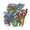
| ||||||||||||||||||||||||||||||||||||||||||||||||
|---|---|---|---|---|---|---|---|---|---|---|---|---|---|---|---|---|---|---|---|---|---|---|---|---|---|---|---|---|---|---|---|---|---|---|---|---|---|---|---|---|---|---|---|---|---|---|---|---|---|
| 1 |
| ||||||||||||||||||||||||||||||||||||||||||||||||
| Unit cell |
| ||||||||||||||||||||||||||||||||||||||||||||||||
| Noncrystallographic symmetry (NCS) | NCS oper:
|
-Components
| #1: Protein | Mass: 16483.488 Da / Num. of mol.: 12 / Mutation: YES Source method: isolated from a genetically manipulated source Source: (gene. exp.) STREPTOMYCES COELICOLOR (bacteria) / Plasmid: PTB-361 / Production host: ESCHERICHIA COLI (E. coli) / Strain (production host): BL21 / References: UniProt: P15474, 3-dehydroquinate dehydratase#2: Chemical | ChemComp-TRS / Tris  Mass: 122.143 Da / Num. of mol.: 4 / Source method: obtained synthetically / Formula: C4H12NO3 / Comment: pH buffer*YM Mass: 122.143 Da / Num. of mol.: 4 / Source method: obtained synthetically / Formula: C4H12NO3 / Comment: pH buffer*YM#3: Chemical | ChemComp-DHK / 3-Dehydroshikimic acid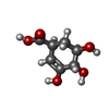  Mass: 174.151 Da / Num. of mol.: 12 / Source method: obtained synthetically / Formula: C7H10O5 Mass: 174.151 Da / Num. of mol.: 12 / Source method: obtained synthetically / Formula: C7H10O5#4: Water | ChemComp-HOH / | Water Mass: 18.015 Da / Num. of mol.: 1580 / Source method: isolated from a natural source / Formula: H2O Mass: 18.015 Da / Num. of mol.: 1580 / Source method: isolated from a natural source / Formula: H2OCompound details | FUNCTION: CATALYSIS VIA AN ENOLATE FOR A TRANS-DEHYDRATIO | |
|---|
-Experimental details
-Experiment
| Experiment | Method: X-RAY DIFFRACTION / Number of used crystals: 1 |
|---|
- Sample preparation
| Crystal | Density Matthews: 2.9 Å3/Da / Density % sol: 56.5 % Description: DATA PROCESSED TO CORNERS OF DETECTOR THEREFORE LOW COMPLETENESS OF HIGHEST RESOLUTION SHELL |
|---|---|
| Crystal grow | pH: 8.5 Details: PEG 8000, SODIUM/POTASSIUM PHOSPHATE, TRIS BUFFER, pH 8.50 |
-Data collection
| Diffraction | Mean temperature: 100 K |
|---|---|
| Diffraction source | Source: SYNCHROTRON / Site: SRS  / Beamline: PX9.6 / Wavelength: 0.87 / Beamline: PX9.6 / Wavelength: 0.87 |
| Detector | Type: ADC QUANTUM 4 / Detector: CCD / Date: Mar 15, 1998 |
| Radiation | Protocol: SINGLE WAVELENGTH / Monochromatic (M) / Laue (L): M / Scattering type: x-ray |
| Radiation wavelength | Wavelength: 0.87 Å / Relative weight: 1 |
| Reflection | Resolution: 1.8→50 Å / Num. obs: 213906 / % possible obs: 98.7 % / Redundancy: 6 % / Rmerge(I) obs: 0.064 |
| Reflection shell | Resolution: 1.6→1.63 Å / % possible all: 21.5 |
- Processing
| Software |
| ||||||||||||||||||||
|---|---|---|---|---|---|---|---|---|---|---|---|---|---|---|---|---|---|---|---|---|---|
| Refinement | Method to determine structure: MOLECULAR REPLACEMENT Starting model: 2DHQ Resolution: 1.6→45 Å / Cross valid method: THROUGHOUT / σ(F): 0 / Details: NONE
| ||||||||||||||||||||
| Refinement step | Cycle: LAST / Resolution: 1.6→45 Å
|
