ムービー
ムービー コントローラー


+ データを開く

- 基本情報
基本情報
| 登録情報 | データベース: PDB / ID: 7tj7 | |||||||||
|---|---|---|---|---|---|---|---|---|---|---|
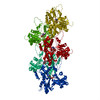
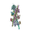
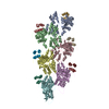
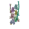
| タイトル | Cardiac thin filament decorated with C1 Ig-domain and regulatory M-domain of cardiac myosin binding protein C (cMyBP-C) | |||||||||
 要素 要素 |
| |||||||||
 キーワード キーワード |  MOTOR PROTEIN (モータータンパク質) / muscle contraction regulator / muscle protein (骨格筋) MOTOR PROTEIN (モータータンパク質) / muscle contraction regulator / muscle protein (骨格筋) | |||||||||
| 機能・相同性 |  機能・相同性情報 機能・相同性情報C zone / regulation of muscle filament sliding / striated muscle myosin thick filament / cardiac myofibril / regulation of striated muscle contraction / Striated Muscle Contraction / positive regulation of ATP-dependent activity / A band / structural constituent of muscle / ventricular cardiac muscle tissue morphogenesis ...C zone / regulation of muscle filament sliding / striated muscle myosin thick filament / cardiac myofibril / regulation of striated muscle contraction / Striated Muscle Contraction / positive regulation of ATP-dependent activity / A band / structural constituent of muscle / ventricular cardiac muscle tissue morphogenesis / myosin binding / myosin heavy chain binding / ATPase activator activity / heart morphogenesis / titin binding / cardiac muscle contraction / sarcomere / actin binding / 細胞接着 / identical protein binding / metal ion binding / 細胞質基質類似検索 - 分子機能 | |||||||||
| 生物種 |  Homo sapiens (ヒト) Homo sapiens (ヒト) Sus scrofa (ブタ) Sus scrofa (ブタ) | |||||||||
| 手法 | 電子顕微鏡法 / らせん対称体再構成法 / クライオ電子顕微鏡法 / 解像度: 8 Å | |||||||||
 データ登録者 データ登録者 | Risi, C.M. / Galkin, V.E. | |||||||||
| 資金援助 |  米国, 2件 米国, 2件
| |||||||||
 引用 引用 | ジャーナル: J Mol Biol / 年: 2022 タイトル: Cryo-Electron Microscopy Reveals Cardiac Myosin Binding Protein-C M-Domain Interactions with the Thin Filament. 著者: Cristina M Risi / Edwin Villanueva / Betty Belknap / Rachel L Sadler / Samantha P Harris / Howard D White / Vitold E Galkin / 要旨: Cardiac myosin binding protein C (cMyBP-C) modulates cardiac contraction via direct interactions with cardiac thick (myosin) and thin (actin) filaments (cTFs). While its C-terminal domains (e.g. C8- ...Cardiac myosin binding protein C (cMyBP-C) modulates cardiac contraction via direct interactions with cardiac thick (myosin) and thin (actin) filaments (cTFs). While its C-terminal domains (e.g. C8-C10) anchor cMyBP-C to the backbone of the thick filament, its N-terminal domains (NTDs) (e.g. C0, C1, M, and C2) bind to both myosin and actin to accomplish its dual roles of inhibiting thick filaments and activating cTFs. While the positions of C0, C1 and C2 on cTF have been reported, the binding site of the M-domain on the surface of the cTF is unknown. Here, we used cryo-EM to reveal that the M-domain interacts with actin via helix 3 of its ordered tri-helix bundle region, while the unstructured part of the M-domain does not maintain extensive interactions with actin. We combined the recently obtained structure of the cTF with the positions of all the four NTDs on its surface to propose a complete model of the NTD binding to the cTF. The model predicts that the interactions of the NTDs with the cTF depend on the activation state of the cTF. At the peak of systole, when bound to the extensively activated cTF, NTDs would inhibit actomyosin interactions. In contrast, at falling Ca levels, NTDs would not compete with the myosin heads for binding to the cTF, but would rather promote formation of active cross-bridges at the adjacent regulatory units located at the opposite cTF strand. Our structural data provides a testable model of the cTF regulation by the cMyBP-C. | |||||||||
| 履歴 |
|
- 構造の表示
構造の表示
| 構造ビューア | 分子: MolmilJmol/JSmol |
|---|
- ダウンロードとリンク
-ダウンロード
| PDBx/mmCIF形式 | 7tj7.cif.gz | 632.7 KB | 表示 | PDBx/mmCIF形式 |
|---|---|---|---|---|
| PDB形式 | pdb7tj7.ent.gz | 517.3 KB | 表示 | PDB形式 |
| PDBx/mmJSON形式 | 7tj7.json.gz | ツリー表示 | PDBx/mmJSON形式 | |
| その他 |  その他のダウンロード その他のダウンロード |
-検証レポート
| アーカイブディレクトリ | https://data.pdbj.org/pub/pdb/validation_reports/tj/7tj7ftp://data.pdbj.org/pub/pdb/validation_reports/tj/7tj7 | HTTPS FTP |
|---|
-関連構造データ


-リンク
 PDBj
PDBj


- 集合体
集合体
| 登録構造単位 | 
|
|---|---|
| 1 |
|
-要素
| #1: タンパク質 | 分子量: 41830.551 Da / 分子数: 6 / 由来タイプ: 天然 / 由来: (天然) Sus scrofa (ブタ) / 器官: heart心臓 / 組織: cardiac muscle / 参照: UniProt: A0A4X1UMF3#2: タンパク質 | 分子量: 24803.123 Da / 分子数: 12 / 由来タイプ: 組換発現 / 由来: (組換発現) Homo sapiens (ヒト) / 遺伝子: MYBPC3 / 発現宿主:  Escherichia coli (大腸菌) / 参照: UniProt: Q14896 Escherichia coli (大腸菌) / 参照: UniProt: Q14896#3: タンパク質 | 分子量: 11507.176 Da / 分子数: 4 / 由来タイプ: 天然 / 由来: (天然) Sus scrofa (ブタ) / 器官: heart心臓 / 組織: cardiac muscle |
|---|
-実験情報
-実験
| 実験 | 手法: 電子顕微鏡法 |
|---|---|
| EM実験 | 試料の集合状態: HELICAL ARRAY / 3次元再構成法: らせん対称体再構成法 |
- 試料調製
試料調製
| 構成要素 | 名称: Cardiac thin filament decorated with C1 Ig-domain and regulatory M-domain of cardiac myosin binding protein C (cMyBP-C) タイプ: COMPLEX 詳細: Thin filaments decorated with C1-M fragment of cMyBP-C show bound triple helix motif of the M-domain and bound C1 Ig-domain. Entity ID: all / 由来: MULTIPLE SOURCES |
|---|---|
| 分子量 | 実験値: NO |
| 緩衝液 | pH: 7 |
| 試料 | 包埋: NO / シャドウイング: NO / 染色: NO / 凍結: YES |
| 試料支持 | グリッドの材料: COPPER / グリッドのサイズ: 300 divisions/in. グリッドのタイプ: PELCO Ultrathin Carbon with Lacey Carbon |
| 急速凍結 | 凍結剤: ETHANE / 湿度: 100 % / 凍結前の試料温度: 275 K |
- 電子顕微鏡撮影
| 実験機器 |  モデル: Titan Krios / 画像提供: FEI Company |
|---|---|
| 顕微鏡 | モデル: FEI TITAN KRIOS |
| 電子銃 | 電子線源: FIELD EMISSION GUN / 加速電圧: 300 kV / 照射モード: FLOOD BEAM |
| 電子レンズ | モード: BRIGHT FIELDBright-field microscopy / 最大 デフォーカス(公称値): 3500 nm / 最小 デフォーカス(公称値): 1000 nm |
| 試料ホルダ | 凍結剤: NITROGEN 試料ホルダーモデル: FEI TITAN KRIOS AUTOGRID HOLDER |
| 撮影 | 電子線照射量: 34 e/Å2 / フィルム・検出器のモデル: GATAN K3 (6k x 4k) |
- 解析
解析
| ソフトウェア | 名称: PHENIX / バージョン: 1.18.2_3874: / 分類: 精密化 | ||||||||||||||||||||||||||||||||
|---|---|---|---|---|---|---|---|---|---|---|---|---|---|---|---|---|---|---|---|---|---|---|---|---|---|---|---|---|---|---|---|---|---|
| EMソフトウェア |
| ||||||||||||||||||||||||||||||||
| CTF補正 | タイプ: PHASE FLIPPING AND AMPLITUDE CORRECTION | ||||||||||||||||||||||||||||||||
| らせん対称 | 回転角度/サブユニット: -166.7 ° / 軸方向距離/サブユニット: 27.4 Å / らせん対称軸の対称性: C1 | ||||||||||||||||||||||||||||||||
| 3次元再構成 | 解像度: 8 Å / 解像度の算出法: FSC 0.143 CUT-OFF / 粒子像の数: 9710 / アルゴリズム: BACK PROJECTION / 対称性のタイプ: HELICAL | ||||||||||||||||||||||||||||||||
| 原子モデル構築 | プロトコル: FLEXIBLE FIT / 空間: REAL | ||||||||||||||||||||||||||||||||
| 原子モデル構築 |
| ||||||||||||||||||||||||||||||||
| 拘束条件 |
|