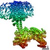
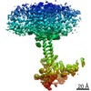
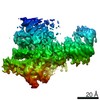
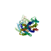
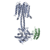
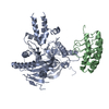
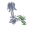
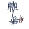
[日本語] English
- PDB-7pdf: focus refinement of soluble domain of adenylyl cyclase 9 in compl... -

+
データを開く


IDまたはキーワード:

読み込み中...

-
基本情報

登録情報
データベース: PDB / ID: 7pdf
タイトルfocus refinement of soluble domain of adenylyl cyclase 9 in complex with Gs protein alpha subunit and MANT-GTP
要素
  • Adenylate cyclase 9アデニル酸シクラーゼ
  • Guanine nucleotide-binding protein G(s) subunit alpha isoforms short
キーワードSIGNALING PROTEIN / membrane protein (膜タンパク質) / adenylyl cyclase (アデニル酸シクラーゼ) / signalling transduction.
機能・相同性
機能・相同性情報


Adenylate cyclase activating pathway / Adenylate cyclase inhibitory pathway / phosphorus-oxygen lyase activity / sensory perception of chemical stimulus / PKA activation / アデニル酸シクラーゼ / mu-type opioid receptor binding / Hedgehog 'off' state / corticotropin-releasing hormone receptor 1 binding / cAMP biosynthetic process ...Adenylate cyclase activating pathway / Adenylate cyclase inhibitory pathway / phosphorus-oxygen lyase activity / sensory perception of chemical stimulus / PKA activation / アデニル酸シクラーゼ / mu-type opioid receptor binding / Hedgehog 'off' state / corticotropin-releasing hormone receptor 1 binding / cAMP biosynthetic process / adenylate cyclase activity / G alpha (z) signalling events / beta-2 adrenergic receptor binding / D1 dopamine receptor binding / positive regulation of cAMP-mediated signaling / adenylate cyclase-activating adrenergic receptor signaling pathway / ionotropic glutamate receptor binding / insulin-like growth factor receptor binding / adenylate cyclase activator activity / G-protein beta/gamma-subunit complex binding / adenylate cyclase-activating G protein-coupled receptor signaling pathway / positive regulation of GTPase activity / adenylate cyclase-activating dopamine receptor signaling pathway / heterotrimeric G-protein complex / in utero embryonic development / intracellular signal transduction / GTPase activity / GTP binding / ATP binding / 生体膜 / metal ion binding / 細胞膜 / 細胞質基質
類似検索 - 分子機能
Adenylyl cyclase class-4/guanylyl cyclase, conserved site / Guanylate cyclase signature. / Adenylyl- / guanylyl cyclase, catalytic domain / Adenylyl cyclase class-3/4/guanylyl cyclase / Adenylate and Guanylate cyclase catalytic domain / Guanylate cyclase domain profile. / Nucleotide cyclase / G-protein alpha subunit, group S / G-alpha domain profile. / Guanine nucleotide binding protein (G-protein), alpha subunit ...Adenylyl cyclase class-4/guanylyl cyclase, conserved site / Guanylate cyclase signature. / Adenylyl- / guanylyl cyclase, catalytic domain / Adenylyl cyclase class-3/4/guanylyl cyclase / Adenylate and Guanylate cyclase catalytic domain / Guanylate cyclase domain profile. / Nucleotide cyclase / G-protein alpha subunit, group S / G-alpha domain profile. / Guanine nucleotide binding protein (G-protein), alpha subunit / G protein alpha subunit, helical insertion / G-protein alpha subunit / G protein alpha subunit / P-loop containing nucleoside triphosphate hydrolase
類似検索 - ドメイン・相同性
5'-GUANOSINE-DIPHOSPHATE-MONOTHIOPHOSPHATE / アデニル酸シクラーゼ / Guanine nucleotide-binding protein G(s) subunit alpha isoforms short
類似検索 - 構成要素
生物種Bos taurus (ウシ)
手法電子顕微鏡法 / 単粒子再構成法 / クライオ電子顕微鏡法 / 解像度: 3.8 Å
データ登録者Qi, C. / Korkhov, V.M.
資金援助2件
組織認可番号
Swiss National Science Foundation150665
Swiss National Science Foundation176992
引用ジャーナル: Nat Commun / : 2022
タイトル: Structural basis of adenylyl cyclase 9 activation.
著者: Chao Qi / Pia Lavriha / Ved Mehta / Basavraj Khanppnavar / Inayathulla Mohammed / Yong Li / Michalis Lazaratos / Jonas V Schaefer / Birgit Dreier / Andreas Plückthun / Ana-Nicoleta Bondar / ...著者: Chao Qi / Pia Lavriha / Ved Mehta / Basavraj Khanppnavar / Inayathulla Mohammed / Yong Li / Michalis Lazaratos / Jonas V Schaefer / Birgit Dreier / Andreas Plückthun / Ana-Nicoleta Bondar / Carmen W Dessauer / Volodymyr M Korkhov /
要旨: Adenylyl cyclase 9 (AC9) is a membrane-bound enzyme that converts ATP into cAMP. The enzyme is weakly activated by forskolin, fully activated by the G protein Gαs subunit and is autoinhibited by the ...Adenylyl cyclase 9 (AC9) is a membrane-bound enzyme that converts ATP into cAMP. The enzyme is weakly activated by forskolin, fully activated by the G protein Gαs subunit and is autoinhibited by the AC9 C-terminus. Although our recent structural studies of the AC9-Gαs complex provided the framework for understanding AC9 autoinhibition, the conformational changes that AC9 undergoes in response to activator binding remains poorly understood. Here, we present the cryo-EM structures of AC9 in several distinct states: (i) AC9 bound to a nucleotide inhibitor MANT-GTP, (ii) bound to an artificial activator (DARPin C4) and MANT-GTP, (iii) bound to DARPin C4 and a nucleotide analogue ATPαS, (iv) bound to Gαs and MANT-GTP. The artificial activator DARPin C4 partially activates AC9 by binding at a site that overlaps with the Gαs binding site. Together with the previously observed occluded and forskolin-bound conformations, structural comparisons of AC9 in the four conformations described here show that secondary structure rearrangements in the region surrounding the forskolin binding site are essential for AC9 activation.
履歴
登録2021年8月5日登録サイト: PDBE / 処理サイト: PDBE
改定 1.02022年1月26日Provider: repository / タイプ: Initial release
改定 1.12022年3月2日Group: Database references / Source and taxonomy / Structure summary
カテゴリ: citation / em_entity_assembly ...citation / em_entity_assembly / em_entity_assembly_naturalsource / em_entity_assembly_recombinant
Item: _citation.country / _citation.journal_abbrev ..._citation.country / _citation.journal_abbrev / _citation.journal_id_CSD / _citation.journal_id_ISSN / _citation.pdbx_database_id_DOI / _citation.title / _citation.year
改定 1.22022年3月9日Group: Database references / カテゴリ: citation / citation_author
Item: _citation.journal_volume / _citation.page_first ..._citation.journal_volume / _citation.page_first / _citation.page_last / _citation.pdbx_database_id_PubMed / _citation.title / _citation_author.identifier_ORCID / _citation_author.name

-
構造の表示

ムービー
  • 登録構造単位
  • Jmolによる作画
  • ダウンロード
  • EMマップとの重ね合わせ
  • マップデータ: EMDB-13336
  • UCSF Chimeraによる作画
  • ダウンロード
ムービービューア
構造ビューア分子:
MolmilJmol/JSmol

ダウンロードとリンク

-
集合体

登録構造単位
A: Adenylate cyclase 9
B: Guanine nucleotide-binding protein G(s) subunit alpha isoforms short
ヘテロ分子


分子量 (理論値)分子数
合計 (水以外)198,7104
ポリマ-198,1472
非ポリマー5642
0
1


  • 登録構造と同一
  • 登録者が定義した集合体
  • 根拠: 電子顕微鏡法
タイプ名称対称操作
identity operation1_5551
Buried area3000 Å2
ΔGint-27 kcal/mol
Surface area36970 Å2

-
要素

#1: タンパク質 Adenylate cyclase 9 / アデニル酸シクラーゼ


分子量: 151142.875 Da / 分子数: 1 / 由来タイプ: 組換発現 / 由来: (組換発現) Bos taurus (ウシ) / 発現宿主: Homo sapiens (ヒト) / 参照: UniProt: E1BM79
#2: タンパク質 Guanine nucleotide-binding protein G(s) subunit alpha isoforms short / Adenylate cyclase-stimulating G alpha protein


分子量: 47003.801 Da / 分子数: 1 / 由来タイプ: 組換発現 / 由来: (組換発現) Bos taurus (ウシ) / 遺伝子: GNAS, GNAS1 / 発現宿主: Trichoplusia ni (イラクサキンウワバ) / 参照: UniProt: P04896
#3: 化合物 ChemComp-GSP / 5'-GUANOSINE-DIPHOSPHATE-MONOTHIOPHOSPHATE / GTP-γ-S


分子量: 539.246 Da / 分子数: 1 / 由来タイプ: 合成 / : C10H16N5O13P3S / タイプ: SUBJECT OF INVESTIGATION
#4: 化合物 ChemComp-MG / MAGNESIUM ION / マグネシウムジカチオン


分子量: 24.305 Da / 分子数: 1 / 由来タイプ: 合成 / : Mg / タイプ: SUBJECT OF INVESTIGATION
研究の焦点であるリガンドがあるかY

-
実験情報

-
実験

実験手法: 電子顕微鏡法
EM実験試料の集合状態: PARTICLE / 3次元再構成法: 単粒子再構成法

-
試料調製

構成要素
ID名称タイプEntity IDParent-ID由来
1Adenylyl cyclase 9 bound to MANT-GTPCOMPLEX#1-#20RECOMBINANT
2Adenylyl cyclase 9アデニル酸シクラーゼCOMPLEX#11RECOMBINANT
3Guanine nucleotide-binding protein G(s) subunit alpha isoforms shortCOMPLEX#21RECOMBINANT
分子量: 146 kDa/nm / 実験値: NO
由来(天然)
IDEntity assembly-ID生物種Ncbi tax-ID
22Bos taurus (ウシ)9913
33Bos taurus (ウシ)9913
由来(組換発現)
IDEntity assembly-ID生物種Ncbi tax-ID細胞
22Homo sapiens (ヒト)9606Human Embryonic Kidney Cells 293
33Trichoplusia ni (イラクサキンウワバ)7111
緩衝液pH: 8
試料濃度: 5 mg/ml / 包埋: NO / シャドウイング: NO / 染色: NO / 凍結: YES
急速凍結凍結剤: ETHANE

-
電子顕微鏡撮影

実験機器
モデル: Titan Krios / 画像提供: FEI Company
顕微鏡モデル: FEI TITAN KRIOS
電子銃電子線源: FIELD EMISSION GUN / 加速電圧: 300 kV / 照射モード: FLOOD BEAM
電子レンズモード: BRIGHT FIELDBright-field microscopy
撮影電子線照射量: 40 e/Å2 / 検出モード: COUNTING
フィルム・検出器のモデル: GATAN K2 SUMMIT (4k x 4k)
電子光学装置エネルギーフィルタースリット幅: 20 eV

-
解析

EMソフトウェア
ID名称カテゴリ
7RELIONモデルフィッティング
9RELIONモデル精密化
CTF補正タイプ: NONE
3次元再構成解像度: 3.8 Å / 解像度の算出法: FSC 0.143 CUT-OFF / 粒子像の数: 157200 / 対称性のタイプ: POINT
原子モデル構築プロトコル: RIGID BODY FIT

+
万見について

-
お知らせ

-
2022年2月9日: EMDBエントリの付随情報ファイルのフォーマットが新しくなりました

EMDBエントリの付随情報ファイルのフォーマットが新しくなりました

  • EMDBのヘッダファイルのバージョン3が、公式のフォーマットとなりました。
  • これまでは公式だったバージョン1.9は、アーカイブから削除されます。

関連情報:EMDBヘッダ

外部リンク:wwPDBはEMDBデータモデルのバージョン3へ移行します

-
2020年8月12日: 新型コロナ情報

新型コロナ情報

URL: https://pdbj.org/emnavi/covid19.php

新ページ: EM Navigatorに新型コロナウイルスの特設ページを開設しました。

関連情報:Covid-19情報 / 2020年3月5日: 新型コロナウイルスの構造データ

+
2020年3月5日: 新型コロナウイルスの構造データ

新型コロナウイルスの構造データ

関連情報:万見生物種 / 2020年8月12日: 新型コロナ情報

外部リンク:COVID-19特集ページ - PDBj / 今月の分子2020年2月:コロナウイルスプロテーアーゼ

+
2019年1月31日: EMDBのIDの桁数の変更

EMDBのIDの桁数の変更

  • EMDBエントリに付与されているアクセスコード(EMDB-ID)は4桁の数字(例、EMD-1234)でしたが、間もなく枯渇します。これまでの4桁のID番号は4桁のまま変更されませんが、4桁の数字を使い切った後に発行されるIDは5桁以上の数字(例、EMD-12345)になります。5桁のIDは2019年の春頃から発行される見通しです。
  • EM Navigator/万見では、接頭語「EMD-」は省略されています。

関連情報:Q: 「EMD」とは何ですか? / 万見/EM NavigatorにおけるID/アクセスコードの表記

外部リンク:EMDB Accession Codes are Changing Soon! / PDBjへお問い合わせ

+
2017年7月12日: PDB大規模アップデート

PDB大規模アップデート

  • 新バージョンのPDBx/mmCIF辞書形式に基づくデータがリリースされました。
  • 今回の更新はバージョン番号が4から5になる大規模なもので、全エントリデータの書き換えが行われる「Remediation」というアップデートに該当します。
  • このバージョンアップで、電子顕微鏡の実験手法に関する多くの項目の書式が改定されました(例:em_softwareなど)。
  • EM NavigatorとYorodumiでも、この改定に基づいた表示内容になります。

外部リンク:wwPDB Remediation / OneDepデータ基準に準拠した、より強化された内容のモデル構造ファイルが、PDBアーカイブで公開されました。

-
万見 (Yorodumi)

幾万の構造データを、幾万の視点から

  • 万見(Yorodumi)は、EMDB/PDB/SASBDBなどの構造データを閲覧するためのページです。
  • EM Navigatorの詳細ページの後継、Omokage検索のフロントエンドも兼ねています。

関連情報:EMDB / PDB / SASBDB / 3つのデータバンクの比較 / 万見検索 / 2016年8月31日: 新しいEM Navigatorと万見 / 万見文献 / Jmol/JSmol / 機能・相同性情報 / 新しいEM Navigatorと万見の変更点

他の情報も見る