 ムービー
ムービー コントローラー


+ データを開く

- 基本情報
基本情報
| 登録情報 | データベース: PDB / ID: 7pck | ||||||
|---|---|---|---|---|---|---|---|
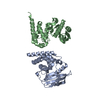
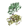
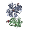
| タイトル | CRYSTAL STRUCTURE OF WILD TYPE HUMAN PROCATHEPSIN K | ||||||
 要素 要素 | PROTEIN (PROCATHEPSIN K) | ||||||
 キーワード キーワード |  HYDROLASE (加水分解酵素) / HYDROLASE (THIOL PROTEASE) / PROCATHEPSIN K / CYSTEINE PROTEASES (システインプロテアーゼ) / PROREGION HYDROLASE (加水分解酵素) / HYDROLASE (THIOL PROTEASE) / PROCATHEPSIN K / CYSTEINE PROTEASES (システインプロテアーゼ) / PROREGION | ||||||
| 機能・相同性 |  機能・相同性情報カテプシンK / mononuclear cell differentiation / intramembranous ossification / negative regulation of cartilage development / cellular response to zinc ion starvation / RUNX1 regulates transcription of genes involved in differentiation of keratinocytes / thyroid hormone generation / endolysosome lumen / Trafficking and processing of endosomal TLR / proteoglycan binding ...カテプシンK / mononuclear cell differentiation / intramembranous ossification / negative regulation of cartilage development / cellular response to zinc ion starvation / RUNX1 regulates transcription of genes involved in differentiation of keratinocytes / thyroid hormone generation / endolysosome lumen / Trafficking and processing of endosomal TLR / proteoglycan binding / Activation of Matrix Metalloproteinases / cysteine-type endopeptidase activator activity involved in apoptotic process / マイトファジー / Collagen degradation / fibronectin binding / collagen catabolic process / extracellular matrix disassembly / cysteine-type peptidase activity / positive regulation of apoptotic signaling pathway / bone resorption / cellular response to transforming growth factor beta stimulus / collagen binding / MHC class II antigen presentation / Degradation of the extracellular matrix / proteolysis involved in protein catabolic process / lysosomal lumen / response to insulin / response to organic cyclic compound / cellular response to tumor necrosis factor / response to ethanol / リソソーム / 免疫応答 / apical plasma membrane / external side of plasma membrane / cysteine-type endopeptidase activity / serine-type endopeptidase activity / intracellular membrane-bounded organelle / タンパク質分解 / extracellular space / extracellular region / 核質 機能・相同性情報カテプシンK / mononuclear cell differentiation / intramembranous ossification / negative regulation of cartilage development / cellular response to zinc ion starvation / RUNX1 regulates transcription of genes involved in differentiation of keratinocytes / thyroid hormone generation / endolysosome lumen / Trafficking and processing of endosomal TLR / proteoglycan binding ...カテプシンK / mononuclear cell differentiation / intramembranous ossification / negative regulation of cartilage development / cellular response to zinc ion starvation / RUNX1 regulates transcription of genes involved in differentiation of keratinocytes / thyroid hormone generation / endolysosome lumen / Trafficking and processing of endosomal TLR / proteoglycan binding / Activation of Matrix Metalloproteinases / cysteine-type endopeptidase activator activity involved in apoptotic process / マイトファジー / Collagen degradation / fibronectin binding / collagen catabolic process / extracellular matrix disassembly / cysteine-type peptidase activity / positive regulation of apoptotic signaling pathway / bone resorption / cellular response to transforming growth factor beta stimulus / collagen binding / MHC class II antigen presentation / Degradation of the extracellular matrix / proteolysis involved in protein catabolic process / lysosomal lumen / response to insulin / response to organic cyclic compound / cellular response to tumor necrosis factor / response to ethanol / リソソーム / 免疫応答 / apical plasma membrane / external side of plasma membrane / cysteine-type endopeptidase activity / serine-type endopeptidase activity / intracellular membrane-bounded organelle / タンパク質分解 / extracellular space / extracellular region / 核質類似検索 - 分子機能 | ||||||
| 生物種 |  Homo sapiens (ヒト) Homo sapiens (ヒト) | ||||||
| 手法 | X線回折 / 分子置換 / 解像度: 3.2 Å | ||||||
 データ登録者 データ登録者 | Sivaraman, J. / Lalumiere, M. / Menard, R. / Cygler, M. | ||||||
 引用 引用 | ジャーナル: Protein Sci. / 年: 1999 タイトル: Crystal structure of wild-type human procathepsin K. 著者: Sivaraman, J. / Lalumiere, M. / Menard, R. / Cygler, M. | ||||||
| 履歴 |
|
- 構造の表示
構造の表示
| 構造ビューア | 分子: MolmilJmol/JSmol |
|---|
- ダウンロードとリンク
-ダウンロード
| PDBx/mmCIF形式 | 7pck.cif.gz | 233 KB | 表示 | PDBx/mmCIF形式 |
|---|---|---|---|---|
| PDB形式 | pdb7pck.ent.gz | 187.3 KB | 表示 | PDB形式 |
| PDBx/mmJSON形式 | 7pck.json.gz | ツリー表示 | PDBx/mmJSON形式 | |
| その他 |  その他のダウンロード その他のダウンロード |
-検証レポート
| アーカイブディレクトリ | https://data.pdbj.org/pub/pdb/validation_reports/pc/7pckftp://data.pdbj.org/pub/pdb/validation_reports/pc/7pck | HTTPS FTP |
|---|
-関連構造データ
| 関連構造データ | 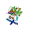 1cjlS S: 精密化の開始モデル |
|---|---|
| 類似構造データ |


-リンク
 PDBj
PDBj


- 集合体
集合体
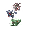
| 登録構造単位 | 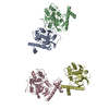
| ||||||||
|---|---|---|---|---|---|---|---|---|---|
| 1 | 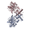
| ||||||||
| 2 | 
| ||||||||
| 単位格子 |
|
-要素
| #1: タンパク質 | 分子量: 35357.898 Da / 分子数: 4 / 由来タイプ: 天然 / 由来: (天然) Homo sapiens (ヒト) / 参照: UniProt: P43235, カテプシンK |
|---|
-実験情報
-実験
| 実験 | 手法: X線回折 / 使用した結晶の数: 2 |
|---|
- 試料調製
試料調製
| 結晶 | マシュー密度: 2.9 Å3/Da / 溶媒含有率: 55 % | ||||||||||||||||||||||||||||||
|---|---|---|---|---|---|---|---|---|---|---|---|---|---|---|---|---|---|---|---|---|---|---|---|---|---|---|---|---|---|---|---|
| 結晶化 | pH: 7 詳細: 100 MM TRIS-HCL, 5% 2-METHYL-2,4-PENTANEDIOL, 12 MM AMMONIUM SULPHATE AND 9% PEG, pH 7.0 | ||||||||||||||||||||||||||||||
| 結晶 | *PLUS | ||||||||||||||||||||||||||||||
| 結晶化 | *PLUS 手法: 蒸気拡散法, ハンギングドロップ法 | ||||||||||||||||||||||||||||||
| 溶液の組成 | *PLUS
|
-データ収集
| 回折 | 平均測定温度: 293 K |
|---|---|
| 放射光源 | 由来: 回転陽極 / タイプ: RIGAKU / 波長: 1.5418 |
| 検出器 | タイプ: MARRESEARCH / 検出器: IMAGE PLATE / 日付: 1997年8月15日 / 詳細: MIRRORS |
| 放射 | モノクロメーター: NI FILTER / プロトコル: SINGLE WAVELENGTH / 単色(M)・ラウエ(L): M / 散乱光タイプ: x-ray |
| 放射波長 | 波長: 1.5418 Å / 相対比: 1 |
| 反射 | 解像度: 3.2→8 Å / Num. obs: 21801 / % possible obs: 87.2 % / Observed criterion σ(I): 1 / 冗長度: 2 % / Rmerge(I) obs: 0.12 / Net I/σ(I): 10 |
| 反射 シェル | 解像度: 3.2→3.3 Å / 冗長度: 2 % / Rmerge(I) obs: 0.26 / Mean I/σ(I) obs: 3 / % possible all: 68 |
| 反射 | *PLUS Num. measured all: 121990 |
- 解析
解析
| ソフトウェア |
| ||||||||||||||||||||||||||||||||||||||||||||||||||||||||||||
|---|---|---|---|---|---|---|---|---|---|---|---|---|---|---|---|---|---|---|---|---|---|---|---|---|---|---|---|---|---|---|---|---|---|---|---|---|---|---|---|---|---|---|---|---|---|---|---|---|---|---|---|---|---|---|---|---|---|---|---|---|---|
| 精密化 | 構造決定の手法: 分子置換 開始モデル: PDB ENTRY 1CJL 解像度: 3.2→8 Å / Data cutoff high absF: 10000000 / Data cutoff low absF: 0.001 / σ(F): 2
| ||||||||||||||||||||||||||||||||||||||||||||||||||||||||||||
| Refine analyze | Luzzati d res low obs: 0 Å | ||||||||||||||||||||||||||||||||||||||||||||||||||||||||||||
| 精密化ステップ | サイクル: LAST / 解像度: 3.2→8 Å
| ||||||||||||||||||||||||||||||||||||||||||||||||||||||||||||
| 拘束条件 |
| ||||||||||||||||||||||||||||||||||||||||||||||||||||||||||||
| Refine LS restraints NCS | NCS model details: RESTRAINTS / Rms dev position: 1.5 Å / Weight position: 300 | ||||||||||||||||||||||||||||||||||||||||||||||||||||||||||||
| ソフトウェア | *PLUS 名称: X-PLOR / バージョン: 3.843 / 分類: refinement | ||||||||||||||||||||||||||||||||||||||||||||||||||||||||||||
| 精密化 | *PLUS 最高解像度: 3.2 Å / 最低解像度: 8 Å / σ(F): 2 / % reflection Rfree: 6 % | ||||||||||||||||||||||||||||||||||||||||||||||||||||||||||||
| 溶媒の処理 | *PLUS | ||||||||||||||||||||||||||||||||||||||||||||||||||||||||||||
| 原子変位パラメータ | *PLUS | ||||||||||||||||||||||||||||||||||||||||||||||||||||||||||||
| 拘束条件 | *PLUS
|
