 Movie
Movie Controller

[English] 日本語
 Yorodumi
- PDB-4ok0: Crystal structure of putative nucleotidyltransferase from H. pylori -
+ Open data

- Basic information
| Entry | Database: PDB / ID: 4ok0 | ||||||
|---|---|---|---|---|---|---|---|
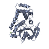
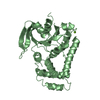
| Title | Crystal structure of putative nucleotidyltransferase from H. pylori | ||||||
 Components Components | Putative | ||||||
 Keywords Keywords | TRANSFERASE / Nucleotidyltransferase | ||||||
| Function / homology |  Function and homology information Function and homology informationJHP933, helical tail domain / JHP933, nucleotidyltransferase-like core domain / Nucleotidyl transferase AbiEii toxin, Type IV TA system / Nucleotidyl transferase AbiEii toxin, Type IV TA system / Nuclear Transport Factor 2; Chain: A, / Methane Monooxygenase Hydroxylase; Chain G, domain 1 / Roll / Up-down Bundle / Mainly Alpha / Alpha Beta Similarity search - Domain/homology | ||||||
| Biological species |   Helicobacter pylori (bacteria) Helicobacter pylori (bacteria) | ||||||
| Method |  X-RAY DIFFRACTION / SYNCHROTRON / SAD / Resolution: 2.17 Å X-RAY DIFFRACTION / SYNCHROTRON / SAD / Resolution: 2.17 Å | ||||||
 Authors Authors | Yoon, J.Y. / Lee, S.J. / Lee, B. / Yang, J.K. / Suh, S.W. | ||||||
 Citation Citation | Journal: Proteins / Year: 2014 Title: Crystal structure of JHP933 from Helicobacter pylori J99 shows two-domain architecture with a DUF1814 family nucleotidyltransferase domain and a helical bundle domain. Authors: Yoon, J.Y. / Lee, S.J. / Kim, D.J. / Lee, B.J. / Yang, J.K. / Suh, S.W. | ||||||
| History |
|
- Structure visualization
| Structure viewer | Molecule: MolmilJmol/JSmol |
|---|
- Downloads & links
-Download
| PDBx/mmCIF format | 4ok0.cif.gz | 105.5 KB | Display | PDBx/mmCIF format |
|---|---|---|---|---|
| PDB format | pdb4ok0.ent.gz | 81.9 KB | Display | PDB format |
| PDBx/mmJSON format | 4ok0.json.gz | Tree view | PDBx/mmJSON format | |
| Others |  Other downloads Other downloads |
-Validation report
| Arichive directory | https://data.pdbj.org/pub/pdb/validation_reports/ok/4ok0ftp://data.pdbj.org/pub/pdb/validation_reports/ok/4ok0 | HTTPS FTP |
|---|
-Related structure data
| Similar structure data |
|---|


-Links
 PDBj
PDBj- Assembly
| Deposited unit | 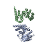
| ||||||||
|---|---|---|---|---|---|---|---|---|---|
| 1 | 
| ||||||||
| 2 | 
| ||||||||
| Unit cell |
|
-Components
| #1: Protein | Mass: 30033.461 Da / Num. of mol.: 2 Source method: isolated from a genetically manipulated source Source: (gene. exp.) Helicobacter pylori (bacteria) / Strain: J99 / ATCC 700824 / Gene: jhp_0933 / Production host: #2: Water | ChemComp-HOH / |  Mass: 18.015 Da / Num. of mol.: 257 / Source method: isolated from a natural source / Formula: H2O Mass: 18.015 Da / Num. of mol.: 257 / Source method: isolated from a natural source / Formula: H2O |
|---|
-Experimental details
-Experiment
| Experiment | Method: X-RAY DIFFRACTION / Number of used crystals: 1 |
|---|
- Sample preparation
| Crystal | Density Matthews: 2.54 Å3/Da / Density % sol: 51.6 % |
|---|---|
| Crystal grow | Temperature: 295 K / Method: vapor diffusion, sitting drop / pH: 7 Details: 200mM ammonium citrate, 20%(w/v) PEG3350, pH 7.0, VAPOR DIFFUSION, SITTING DROP, temperature 295K |
-Data collection
| Diffraction | Mean temperature: 100 K |
|---|---|
| Diffraction source | Source: SYNCHROTRON / Type: OTHER / Wavelength: 0.99999 Å |
| Detector | Type: ADSC QUANTUM 315 / Detector: CCD / Date: Feb 8, 2010 |
| Radiation | Protocol: SINGLE WAVELENGTH / Monochromatic (M) / Laue (L): M / Scattering type: x-ray |
| Radiation wavelength | Wavelength: 0.99999 Å / Relative weight: 1 |
| Reflection | Resolution: 2.17→20 Å / Num. obs: 31472 / % possible obs: 100 % / Observed criterion σ(F): 0 / Observed criterion σ(I): -3 |
| Reflection shell | Resolution: 2.17→2.25 Å / % possible all: 100 |
- Processing
| Software |
| |||||||||||||||||||||||||||||||||||||||||||||
|---|---|---|---|---|---|---|---|---|---|---|---|---|---|---|---|---|---|---|---|---|---|---|---|---|---|---|---|---|---|---|---|---|---|---|---|---|---|---|---|---|---|---|---|---|---|---|
| Refinement | Method to determine structure: SAD / Resolution: 2.17→20 Å / Cor.coef. Fo:Fc: 0.957 / Cor.coef. Fo:Fc free: 0.93 / SU B: 5.105 / SU ML: 0.13 / Cross valid method: THROUGHOUT / ESU R: 0.222 / ESU R Free: 0.192 / Stereochemistry target values: MAXIMUM LIKELIHOOD / Details: HYDROGENS HAVE BEEN USED IF PRESENT IN THE INPUT
| |||||||||||||||||||||||||||||||||||||||||||||
| Solvent computation | Ion probe radii: 0.8 Å / Shrinkage radii: 0.8 Å / VDW probe radii: 1.2 Å / Solvent model: MASK | |||||||||||||||||||||||||||||||||||||||||||||
| Displacement parameters | Biso mean: 36.867 Å2
| |||||||||||||||||||||||||||||||||||||||||||||
| Refinement step | Cycle: LAST / Resolution: 2.17→20 Å
| |||||||||||||||||||||||||||||||||||||||||||||
| Refine LS restraints |
| |||||||||||||||||||||||||||||||||||||||||||||
| LS refinement shell | Resolution: 2.17→2.228 Å / Total num. of bins used: 20
|
