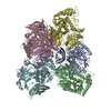
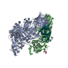
登録情報 データベース : PDB / ID : 3nvqタイトル Molecular mechanism of guidance cue recognition キーワード / / / 機能・相同性 分子機能 ドメイン・相同性 構成要素
/ / / / / / / / / / / / / / / / / / / / / / / / / / / / / / / / / / / / / / / / / / / / / / / / / / / / / / / / / / / / / / / / / / / / / / / / / / / / / / / / / / / / / / / / / / / / 生物種 Homo sapiens (ヒト)手法 / / 解像度 : 2.4 Å データ登録者 Juo, Z. / Liu, H. / Shim, A. / Focia, P. / Chen, X. / Garcia, C. / He, X. ジャーナル : Cell(Cambridge,Mass.) / 年 : 2010タイトル : Structural Basis of Semaphorin-Plexin Recognition and Viral Mimicry from Sema7A and A39R Complexes with PlexinC1.著者 : Liu, H. / Juo, Z.S. / Shim, A.H. / Focia, P.J. / Chen, X. / Garcia, K.C. / He, X. 履歴 登録 2010年7月8日 登録サイト / 処理サイト 改定 1.0 2010年9月15日 Provider / タイプ 改定 1.1 2011年7月13日 Group 改定 2.0 2020年7月29日 Group Advisory / Atomic model ... Advisory / Atomic model / Data collection / Database references / Derived calculations / Structure summary カテゴリ atom_site / chem_comp ... atom_site / chem_comp / database_PDB_caveat / entity / pdbx_chem_comp_identifier / pdbx_entity_nonpoly / pdbx_unobs_or_zero_occ_atoms / struct_conn / struct_ref_seq_dif / struct_site / struct_site_gen Item _atom_site.auth_atom_id / _atom_site.label_atom_id ... _atom_site.auth_atom_id / _atom_site.label_atom_id / _chem_comp.name / _chem_comp.type / _entity.pdbx_description / _pdbx_entity_nonpoly.name / _pdbx_unobs_or_zero_occ_atoms.auth_atom_id / _pdbx_unobs_or_zero_occ_atoms.label_atom_id / _struct_conn.pdbx_dist_value / _struct_conn.pdbx_leaving_atom_flag / _struct_conn.pdbx_role / _struct_conn.ptnr1_auth_asym_id / _struct_conn.ptnr1_auth_comp_id / _struct_conn.ptnr1_auth_seq_id / _struct_conn.ptnr1_label_asym_id / _struct_conn.ptnr1_label_atom_id / _struct_conn.ptnr1_label_comp_id / _struct_conn.ptnr1_label_seq_id / _struct_conn.ptnr2_auth_asym_id / _struct_conn.ptnr2_auth_comp_id / _struct_conn.ptnr2_auth_seq_id / _struct_conn.ptnr2_label_asym_id / _struct_conn.ptnr2_label_atom_id / _struct_conn.ptnr2_label_comp_id / _struct_conn.ptnr2_label_seq_id / _struct_ref_seq_dif.details 解説 / Provider / タイプ
すべて表示 表示を減らす
 ムービー
ムービー コントローラー


 データを開く
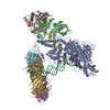
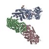
 基本情報
基本情報 要素
要素 キーワード
キーワード 機能・相同性情報
 Homo sapiens (ヒト)
 X線回折 /
  データ登録者
 引用
引用 構造の表示
構造の表示 ダウンロードとリンク
 その他のダウンロード
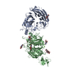


 PDBj
PDBj


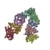
 集合体
集合体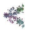

 タイプ: D-saccharide, beta linking / 分子量: 221.208 Da / 分子数: 21 / 由来タイプ: 組換発現 / 式: C8H15NO6
 タイプ: D-saccharide, alpha linking / 分子量: 221.208 Da / 分子数: 1 / 由来タイプ: 組換発現 / 式: C8H15NO6
 分子量: 18.015 Da / 分子数: 858 / 由来タイプ: 天然 / 式: H2O
 試料調製
試料調製 / ビームライン: 21-ID-D / 波長: 1.5418 Å
 解析
解析