| 登録情報 | データベース: PDB / ID: 1i7i
|
|---|
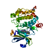
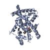
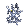
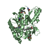
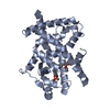
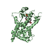
| タイトル | CRYSTAL STRUCTURE OF THE LIGAND BINDING DOMAIN OF HUMAN PPAR-GAMMA IN COMPLEX WITH THE AGONIST AZ 242 |
|---|
 要素 要素 | PEROXISOME PROLIFERATOR ACTIVATED RECEPTOR GAMMA |
|---|
 キーワード キーワード |  TRANSCRIPTION (転写 (生物学)) / anti parallel helix sandwich TRANSCRIPTION (転写 (生物学)) / anti parallel helix sandwich |
|---|
| 機能・相同性 |  機能・相同性情報 機能・相同性情報
prostaglandin receptor activity / regulation of cholesterol transporter activity / negative regulation of connective tissue replacement involved in inflammatory response wound healing / negative regulation of receptor signaling pathway via STAT / MECP2 regulates transcription factors / negative regulation of extracellular matrix assembly / negative regulation of vascular endothelial cell proliferation / negative regulation of cellular response to transforming growth factor beta stimulus / negative regulation of cardiac muscle hypertrophy in response to stress / arachidonic acid binding ...prostaglandin receptor activity / regulation of cholesterol transporter activity / negative regulation of connective tissue replacement involved in inflammatory response wound healing / negative regulation of receptor signaling pathway via STAT / MECP2 regulates transcription factors / negative regulation of extracellular matrix assembly / negative regulation of vascular endothelial cell proliferation / negative regulation of cellular response to transforming growth factor beta stimulus / negative regulation of cardiac muscle hypertrophy in response to stress / arachidonic acid binding / positive regulation of low-density lipoprotein receptor activity / positive regulation of adiponectin secretion / lipoprotein transport / negative regulation of sequestering of triglyceride / positive regulation of vascular associated smooth muscle cell apoptotic process / macrophage derived foam cell differentiation / DNA binding domain binding / STAT family protein binding / positive regulation of fatty acid metabolic process / response to lipid / negative regulation of SMAD protein signal transduction / LBD domain binding / negative regulation of type II interferon-mediated signaling pathway / negative regulation of cholesterol storage / E-box binding / alpha-actinin binding / negative regulation of blood vessel endothelial cell migration / lipid homeostasis / negative regulation of vascular associated smooth muscle cell proliferation / R-SMAD binding / monocyte differentiation / positive regulation of cholesterol efflux / negative regulation of macrophage derived foam cell differentiation / cellular response to low-density lipoprotein particle stimulus / negative regulation of lipid storage / negative regulation of BMP signaling pathway / white fat cell differentiation / negative regulation of mitochondrial fission / positive regulation of fat cell differentiation / negative regulation of osteoblast differentiation / long-chain fatty acid transport / retinoic acid receptor signaling pathway / positive regulation of DNA binding / 細胞分化 / BMP signaling pathway / nuclear retinoid X receptor binding / negative regulation of signaling receptor activity / regulation of cellular response to insulin stimulus / cell maturation / positive regulation of adipose tissue development / epithelial cell differentiation / peroxisome proliferator activated receptor signaling pathway / hormone-mediated signaling pathway / negative regulation of angiogenesis / response to nutrient / negative regulation of MAP kinase activity / fatty acid metabolic process / negative regulation of miRNA transcription / placenta development / Regulation of PTEN gene transcription / negative regulation of smooth muscle cell proliferation / transcription coregulator binding / peptide binding / negative regulation of transforming growth factor beta receptor signaling pathway / SUMOylation of intracellular receptors / mRNA transcription by RNA polymerase II / lipid metabolic process / regulation of circadian rhythm / PPARA activates gene expression / negative regulation of inflammatory response / DNA-binding transcription repressor activity, RNA polymerase II-specific / 血圧 / Transcriptional regulation of white adipocyte differentiation / positive regulation of miRNA transcription / Nuclear Receptor transcription pathway / RNA polymerase II transcription regulator complex / cellular response to insulin stimulus / activation of cysteine-type endopeptidase activity involved in apoptotic process / : / rhythmic process / nuclear receptor activity / positive regulation of DNA-binding transcription factor activity / glucose homeostasis / cellular response to hypoxia / double-stranded DNA binding / DNA-binding transcription activator activity, RNA polymerase II-specific / DNA-binding transcription factor binding / sequence-specific DNA binding / nucleic acid binding / 細胞分化 / receptor complex / transcription cis-regulatory region binding / DNA-binding transcription factor activity, RNA polymerase II-specific / RNA polymerase II cis-regulatory region sequence-specific DNA binding / DNA-binding transcription factor activity / negative regulation of gene expression / 自然免疫系 / intracellular membrane-bounded organelle / negative regulation of DNA-templated transcription / chromatin binding類似検索 - 分子機能 PPARγ / Peroxisome proliferator-activated receptor gamma, N-terminal / PPAR gamma N-terminal region / PPAR / Retinoid X Receptor / Retinoid X Receptor / 核内受容体 / Nuclear hormones receptors DNA-binding region signature. / Zinc finger, nuclear hormone receptor-type / Zinc finger, C4 type (two domains) ...PPARγ / Peroxisome proliferator-activated receptor gamma, N-terminal / PPAR gamma N-terminal region / PPAR / Retinoid X Receptor / Retinoid X Receptor / 核内受容体 / Nuclear hormones receptors DNA-binding region signature. / Zinc finger, nuclear hormone receptor-type / Zinc finger, C4 type (two domains) / Nuclear hormone receptors DNA-binding domain profile. / c4 zinc finger in nuclear hormone receptors / Nuclear hormone receptor, ligand-binding domain / Nuclear hormone receptor-like domain superfamily / Ligand-binding domain of nuclear hormone receptor / Nuclear receptor (NR) ligand-binding (LBD) domain profile. / Ligand binding domain of hormone receptors / Zinc finger, NHR/GATA-type / Orthogonal Bundle / Mainly Alpha類似検索 - ドメイン・相同性 |
|---|
| 生物種 |  Homo sapiens (ヒト) Homo sapiens (ヒト) |
|---|
| 手法 | X線回折 / 分子置換 / 解像度: 2.35 Å |
|---|
 データ登録者 データ登録者 | Petersen, J.F.W. / Cronet, P. / Folmer, R. / Blomberg, N. / Sjoblom, K. / Karlsson, U. / Lindstedt, E.-L. / Bamberg, K. |
|---|
 引用 引用 | ジャーナル: Structure / 年: 2001
タイトル: Structure of the PPARalpha and -gamma ligand binding domain in complex with AZ 242; ligand selectivity and agonist activation in the PPAR family.
著者: Cronet, P. / Petersen, J.F. / Folmer, R. / Blomberg, N. / Sjoblom, K. / Karlsson, U. / Lindstedt, E.L. / Bamberg, K. |
|---|
| 履歴 | | 登録 | 2001年3月9日 | 登録サイト: RCSB / 処理サイト: RCSB |
|---|
| 改定 1.0 | 2002年3月9日 | Provider: repository / タイプ: Initial release |
|---|
| 改定 1.1 | 2008年4月27日 | Group: Version format compliance |
|---|
| 改定 1.2 | 2011年7月13日 | Group: Version format compliance |
|---|
| 改定 1.3 | 2023年8月9日 | Group: Data collection / Database references ...Data collection / Database references / Derived calculations / Refinement description
カテゴリ: chem_comp_atom / chem_comp_bond ...chem_comp_atom / chem_comp_bond / database_2 / pdbx_initial_refinement_model / struct_ref_seq_dif / struct_site
Item: _database_2.pdbx_DOI / _database_2.pdbx_database_accession ..._database_2.pdbx_DOI / _database_2.pdbx_database_accession / _struct_ref_seq_dif.details / _struct_site.pdbx_auth_asym_id / _struct_site.pdbx_auth_comp_id / _struct_site.pdbx_auth_seq_id |
|---|
|
|---|
 ムービー
ムービー コントローラー


 データを開く
 基本情報
基本情報 構造の表示
構造の表示 ダウンロードとリンク
 その他のダウンロード
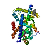
 PDBj
PDBj 集合体
集合体

 分子量: 408.465 Da / 分子数: 2 / 由来タイプ: 合成 / 式: C20H24O7S / コメント: アゴニスト*YM
 分子量: 18.015 Da / 分子数: 62 / 由来タイプ: 天然 / 式: H2O
 試料調製
試料調製 解析
解析